. 2008 Jun 15;586(12):2841-53.
doi: 10.1113/jphysiol.2008.151027. Epub 2008 Apr 17.

Phosphocreatine as an energy source for actin cytoskeletal rearrangements during myoblast fusion

Affiliations

Phosphocreatine as an energy source for actin cytoskeletal rearrangements during myoblast fusion

Roddy S O'Connor et al. J Physiol. .

Abstract

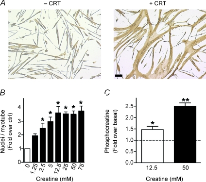
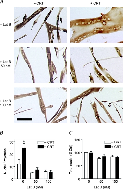
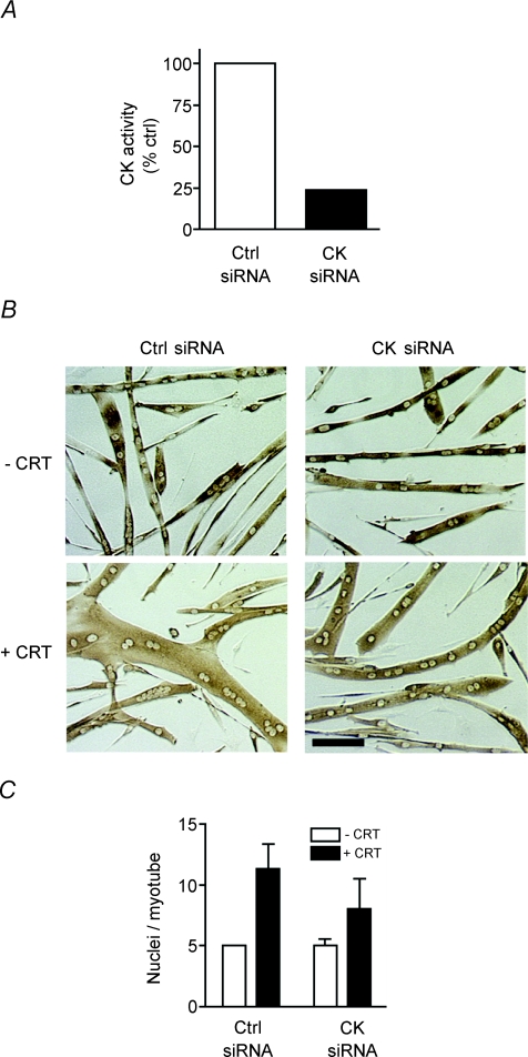
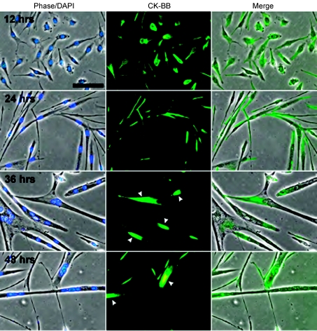
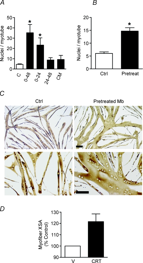
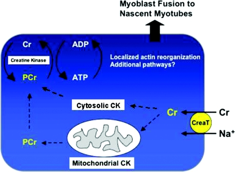
Myoblast fusion is essential for muscle development, postnatal growth and muscle repair after injury. Recent studies have demonstrated roles for actin polymerization during myoblast fusion. Dynamic cytoskeletal assemblies directing cell-cell contact, membrane coalescence and ultimately fusion require substantial cellular energy demands. Various energy generating systems exist in cells but the partitioning of energy sources during myoblast fusion is unknown. Here, we demonstrate a novel role for phosphocreatine (PCr) as a spatiotemporal energy buffer during primary mouse myoblast fusion with nascent myotubes. Creatine treatment enhanced cell fusion in a creatine kinase (CK)-dependent manner suggesting that ATP-consuming reactions are replenished through the PCr/CK system. Furthermore, selective inhibition of actin polymerization prevented myonuclear addition following creatine treatment. As myotube formation is dependent on cytoskeletal reorganization, our findings suggest that PCr hydrolysis is coupled to actin dynamics during myoblast fusion. We conclude that myoblast fusion is a high-energy process, and can be enhanced by PCr buffering of energy demands during actin cytoskeletal rearrangements in myoblast fusion. These findings implicate roles for PCr as a high-energy phosphate buffer in the fusion of multiple cell types including sperm/oocyte, trophoblasts and macrophages. Furthermore, our results suggest the observed beneficial effects of oral creatine supplementation in humans may result in part from enhanced myoblast fusion.


Figures

Figure 1
Figure 1. Creatine enhances myonuclear addition in a dose-dependent manner
A, representative images of primary mouse muscle cells differentiated for 48 h in the absence (–) or presence (+) of 75 mm creatine (CRT) are shown. Bar: 100 μm. B, a dose-dependent increase occurred in myonuclear number with creatine treatment for 48 h with evidence of saturation at higher concentrations. Data are means ±s.e.m. from three independent experiments. (*P < 0.05 relative to control.) C, primary myoblasts were treated with either 12.5 or 50 mm creatine. Creatine enhanced intracellular PCr accumulation as measured by HPLC analysis. All data were normalized to protein content and values are means ±s.e.m. from three independent experiments. (*P < 0.05 relative to control.)
Figure 2
Figure 2. Actin polymerization is required for creatine-mediated effects
A, myoblasts were differentiated in the presence or absence of 12.5 mm creatine (CRT) for 24 h. This medium was subsequently replaced with DM containing vehicle, 50 or 100 nm latrunculin B (Lat B) for the remainder of myogenesis. Representative images following 48 h of differentiation are shown. Bar: 100 μm. B, myonuclear number was reduced to control levels in cells that received either 50 or 100 nm Lat B following creatine treatment. C, no significant difference in the total number of nuclei per field occurred with either creatine or Lat B treatment. Data are means ±s.e.m. from three independent experiments. (P < 0.05.)
Figure 3
Figure 3. Myoblast motility is unaffected by creatine
A, myoblasts were differentiated in the presence and absence of 12.5 mm creatine (CRT) for approximately 24 h and then time-lapse photographs were recorded every 6 min for 80 min. The migratory paths of individual mononucleated cells are shown. Paths of 8–10 cells from each of three independent experiments were pooled for a combined total of 26 paths. B, the mean velocity was similar in control and creatine-treated cells. Data are scatter plots of 48 cells (approximately 15 cells per experiment).
Figure 4
Figure 4. Creatine enhances myoblast fusion independent of proliferation or differentiation
Myoblasts were differentiated in the presence and absence of 75 mm creatine (CRT) for either 24 or 48 h and subsequently immunostained for eMyHC. A, no significant difference in the total number of nuclei per field occurred with creatine treatment. B, creatine did not effect the percentage of nuclei in eMyHC+ cells at 24 h. C, the fusion index was significantly increased in cells treated with creatine for 48 h. D, the number of myotubes per field was significantly decreased following 48 h of creatine treatment. Data are means ±s.e.m. from three independent experiments. (*P < 0.05.)
Figure 5
Figure 5. Creatine enhances myonuclear addition through a CK-dependent mechanism
A, myoblasts containing siRNA for CKB and CKM or a control siRNA were differentiated for 42–46 h. Myotube lysates were analysed for creatine kinase (CK) activity. Data are means from two independent experiments. B, representative images of siRNA containing cells following differentiation for 48 h in the absence (–CRT) and presence of 12.5 mm creatine (+CRT) are shown. Bar: 100 μm. C, following creatine treatment myonuclear number was decreased in cells containing siRNA for CK. Data are means ±s.e.m. from three independent experiments.
Figure 6
Figure 6. CK-BB becomes highly localized as myoblast differentiation and fusion progresses
Representative images obtained from myoblasts differentiated for 12, 24, 36 and 48 h and immunostained with a CK-BB antibody. The nuclei were visualized with DAPI. After 12 h in DM, CK-BB was fairly regularly distributed throughout the cytoplasm but became prominently localized to the ends of myotubes after 36–48 h (arrowheads). Bar = 40 μm.
Figure 7
Figure 7. Pretreating proliferating myoblasts with creatine increases myonuclear addition to nascent myotubes
A, myoblasts were differentiated for 48 h in the presence or absence (C) of 75 mm creatine in DM. 0–48: cells received creatine throughout myogenesis. 0–24: cells received creatine during the first 24 h of differentiation only. This medium was subsequently replaced with DM for the later 24 h of myogenesis. 24–48: these cells received DM for the initial 24 h of differentiation. These cells were supplemented with 75 mm creatine without a media change from 24 to 48 h. CM: cells received DM for the initial 24 h of differentiation. This was replaced with conditioned medium (CM) collected from a parallel dish of differentiating myoblasts treated with creatine during the initial 24 h of myogenesis. Data are means ±s.e.m. from three independent experiments. (*P < 0.05 relative to control.) No statistical difference was observed between 0–24 h and 0–48 h of creatine treatment. B, proliferating myoblasts were cultured in GM containing 12.5 mm creatine for 2 days and then switched to DM in the absence of creatine for 48 h. Myonuclear number was significantly increased in cells that received creatine prior to the induction of differentiation. Data are means ±s.e.m. from three independent experiments. (*P < 0.05 relative to control.) C, representative images of cells pretreated with creatine as myoblasts (Mb) are shown. Bar: 100 μm. D, the XSA of regenerating TA myofibres 5 days following injury was increased in creatine-treated mice. Values are means ±s.e.m., n = 9 per treatment group.
Figure 8
Figure 8. Working model for enhanced myonuclear addition following creatine treatment
Similar to other cells, creatine is transported into muscle cells through the creatine transporter (CreaT). Subsequently, creatine is converted to PCr by mitochondrial and cytosolic CK. We propose that PCr diffuses to sites of high-energy demands where it acts as a spatiotemporal energy buffer during myoblast fusion. PCr hydrolysis by local CKB or CKM provides dynamic bursts of energy during actin cytoskeletal rearrangements associated with the fusion of myoblasts to nascent myotubes.

Comment in

References

    1. Abbott KL, Friday BB, Thaloor D, Murphy TJ, Pavlath GK. Activation and cellular localization of the cyclosporine A-sensitive transcription factor NF-AT in skeletal muscle cells. Mol Biol Cell. 1998;9:2905–2916. - PMC - PubMed
    1. Alfieri RR, Bonelli MA, Cavazzoni A, Brigotti M, Fumarola C, Sestili P, Mozzoni P, De Palma G, Mutti A, Carnicelli D, Vacondio F, Silva C, Borghetti AF, Wheeler KP, Petronini PG. Creatine as a compatible osmolyte in muscle cells exposed to hypertonic stress. J Physiol. 2006;576:391–401. - PMC - PubMed
    1. Ayscough KR, Stryker J, Pokala N, Sanders M, Crews P, Drubin DG. High rates of actin filament turnover in budding yeast and roles for actin in establishment and maintenance of cell polarity revealed using the actin inhibitor latrunculin-A. J Cell Biol. 1997;137:399–416. - PMC - PubMed
    1. Belmont LD, Patterson GM, Drubin DG. New actin mutants allow further characterization of the nucleotide binding cleft and drug binding sites. J Cell Sci. 1999;112:1325–1336. - PubMed
    1. Bernstein BW, Bamburg JR. Actin-ATP hydrolysis is a major energy drain for neurons. J Neurosci. 2003;23:1–6. - PMC - PubMed

Publication types