. 2008 Aug;149(8):4095-105.
doi: 10.1210/en.2008-0249. Epub 2008 Apr 17.

Prolactin promotes the secretion of active cathepsin D at the basal side of rat mammary acini

Affiliations

Prolactin promotes the secretion of active cathepsin D at the basal side of rat mammary acini

Roberta Castino et al. Endocrinology. 2008 Aug.

Abstract

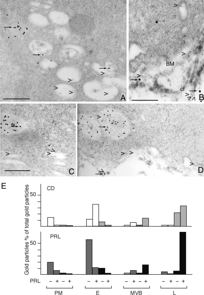
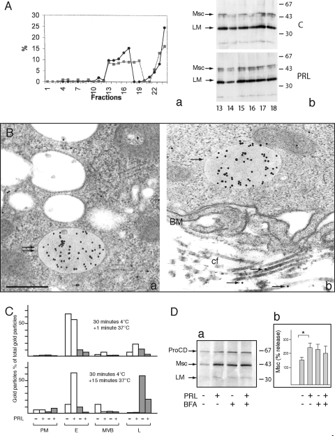
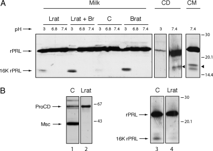
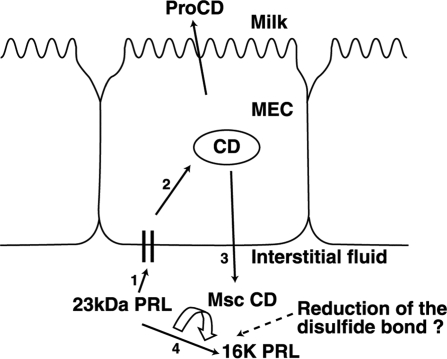
Cathepsin D (CD), a lysosomal aspartic protease present in mammary tissue and milk in various molecular forms, is also found in the incubation medium of mammary acini in molecular forms that are proteolytically active on prolactin at a physiological pH. Because prolactin controls the vesicular traffic in mammary cells, we studied, in vivo and in vitro, its effects on the polarized transport and secretion of various forms of CD in the rat mammary gland. CD accumulated in vesicles not involved in endocytosis in the basal region of cells. Prolactin increased this accumulation and the release of endosomal active single-chain CD at the basal side of acini. The CD-mediated proteolysis of prolactin, leading to the antiangiogenic 16-kDa form, at a physiological pH, was observed only in conditioned medium but not milk. These data support the novel concept that an active molecular form of CD, secreted at the basal side of the mammary epithelium, participates in processing blood-borne prolactin outside the cell, this polarized secretion being controlled by prolactin itself.


Figures

Figure 1
Figure 1
Characterization of CD in serum, milk, conditioned medium, and mammary tissues, from Brat and Lrat. A, Proteins from serum, conditioned medium (CM), and skim milk were analyzed by SDS-PAGE and treated for immunoblotting, as described in Materials and Methods. Serum samples were precipitated by Pst-agarose (Pst.A) before analysis. Lane 1, Serum from lactating rat. Lane 2, Serum from Brat treated for 24 h. Lane 3, Serum from Lrat. Pro-CD was revealed in blood serum from control, Brats, and Lrats. Lane 4, Molecular forms of CD present in the conditioned medium (CM). Lanes 5–8, Molecular forms detected in skim milk. In skim milk from control rats, pro-CD was the principal CD form found in samples that had been previously precipitated (lane 8) or not (lane 6) by Pst-agarose. In skim milk from Lrat (lane 5) and Brat (lane 7), a CD-immunoreactive band with an apparent molecular mass of 46 kDa is prominent. This form was faintly detectable in skim milk from control rat (lanes 6 and 8). Position of the pro-CD, Msc, and LM is on the right. Position of the molecular mass markers (kilodaltons) on the left. B, Immunofluorescence localization of CD in mammary acini. a, Mammary acini of lactating rat. CD was strongly accumulated in the basal region of the acini (arrows) and was present as spots in the cytoplasm (arrowhead). b, After a 24-h bromocriptine-treatment, CD labeling in the basal region of acini was attenuated (arrow). CD was present as spots in the cytoplasm (arrowheads). c, After a 48-h bromocriptine treatment, CD labeling was diffuse in the cytoplasm and accumulated in the lumen. Acinar lumen (asterisk). Bar, 20 μm.
Figure 2
Figure 2
Immunogold electron microscopy localization of CD in the mammary epithelial cells of lactating rat mammary tissue. A, CD was localized in different vesicles (arrows). SV, Secretory vesicle. B, In the basal region of the epithelium, dense vesicles (double arrow) and clear vesicles (arrows) were labeled. C, In the Golgi region, CD was present in Golgi saccules (GS) and frequently associated with coated pits and vesicles budding of the TGN (arrows). Bar, 1 μm.
Figure 3
Figure 3
ConA and CD do not localize in the same vesicles in the basal region of lactating rat mammary epithelial cells. Mammary acini were incubated for 30 min at 4 C and then chased for 5 min at 37 C in the presence of FITC-conjugated conA (arrowheads) (A) or gold-conjugated conA 5-nm gold particles (arrowheads) (C), fixed, and treated for either immunofluorescence detection of CD with antirat CD antibody and FITC-conjugated secondary antibody (arrows) (B) or for the immunogold electron microscopy detection of CD with antirat CD antibody and with a secondary antibody conjugated to 15 nm colloidal gold (arrows) (C). A, ConA strongly stained the periphery of cells. B, CD was observed as spots in the cytoplasm and at the basal region of cells. C, Immunoelectron microscopy shows that gold-conjugated conA (5 nm gold particles; arrowhead) are present in vesicles that differ from those labeled for CD (15 nm gold particles, arrow). BM, Basal membrane. Bar (A and B), 20 μm, (C), 200 nm.
Figure 4
Figure 4
Distribution of CD in vesicles labeled or not by cationized ferritin. Mammary acini were incubated for 30 min at 4 C in Hanks’ medium containing 1 mg/ml cationized ferritin and then incubated at 37 C for 1 min in the absence (A) or presence of 1 μg/ml PRL (B). A, Cationized ferritin was associated with patches of the basal membrane (BM) and was detected in a vesicular tubular network close to the membrane (arrowheads). B, After incubation in the presence of PRL, cationized ferritin was frequently accumulated in cytoplasmic vesicles close to the Golgi apparatus (arrowhead). C, Double localization of CD and cationized ferritin in mammary acini incubated for 30 min at 4 C, chased, and then treated for the immunogold detection of CD, as described above. Cationized ferritin (arrowheads) and CD (arrow) were accumulated separately in different vesicles, whereas a lysosome contained both markers (arrow and arrowhead). Bar, 200 nm. D, Evolution of CD distribution in the different types of vesicles localized in the third basal part of mammary cells. The number of gold particles in percentage of the total gold particles was evaluated in vesicles not labeled with cationized ferritin (white bar) and in vesicles containing cationized ferritin (gray bar). PM, Plasma membrane; E, endosome-like vesicles; MVB, multivesicular bodies; L, lysosomes; right and left panel, means ± sem, two rats; central panel, one rat.
Figure 5
Figure 5
Double localization of CD and PRL in mammary epithelial cells before (A and B) and after (C and D) incubation in the presence of PRL. A and B, Mammary tissue from lactating rat was fixed and treated for immunoelectron microscopy, first on one side of the grid with the anti-CD antibody, followed by 15 nm gold-conjugated antirabbit IgG, and then on the other side of the grid, with the anti-PRL antibody followed by the 5-nm gold-conjugated antirabbit IgG. A, PRL was detected in Golgi vesicles and secretory vesicles (arrowheads); CD was detected in dense vesicles and Golgi saccules (arrows). Bar, 200 nm. B, Endosomes close to the basal membrane (BM) contained PRL alone (arrowhead). CD (arrows) and PRL (arrowheads) were present in the extracellular matrix and associated with collagen filaments (cf.). Bar, 100 nm. C and D, Acini were incubated for 15 min in the presence of 1 μg/ml PRL, fixed, and treated for immunogold electron microscopy with anti-CD antibody and anti-PRL antibody as above. C, CD was accumulated in a vesicle close to the basal membrane (arrow) and rPRL was detected in different vesicles (arrowheads). D, CD and PRL were present in the extracellular matrix close to the basal membrane. An accumulation of PRL and CD were visible in lysosomes characterized by their heterogeneous content (arrow and arrowhead). Bar, 200 nm. E, Cellular distribution of CD (upper panel) and PRL (lower panel) in different cellular compartments of acini incubated in the presence or absence of PRL as described above. Results from one experiment representative of two experiments (20 micrographs were counted for each treatment). The gold particles (5 and 15 nm) corresponding to PRL and CD, respectively, were counted separately on the plasma membrane (PM) and the different vesicles. E, Endosome-like vesicles; MVB, multivesicular bodies; Lys, lysosomes. White bars, percentage of gold particles corresponding to CD in vesicles containing only CD. Clear gray bars, Percentage of gold particles corresponding to CD in vesicles containing both CD and PRL; gray bars, percentage of gold particles corresponding to PRL in vesicles containing only PRL; black bars, percentage of gold particles corresponding to PRL in vesicles containing both gold particles.
Figure 6
Figure 6
In vitro effect of PRL on the intracellular transport and secretion of CD in lactating rat mammary epithelial cells. A, Mammary tissues were incubated for 15 min in the absence or presence of 1 μg/ml PRL, homogenized, and cell fractionated. The content of lysosomal β-hexosaminidase was evaluated in fractions from control mammary tissue (gray squares) and PRL-treated mammary tissues (black spots) (a). Molecular forms of CD accumulating in cell fractions enriched with lysosomal β-hexosaminidase (fractions 13–18) were identified by immunoblotting (b). The mature LM of the double chain of CD is the most represented form. In the presence of PRL, the immunoreactive band corresponding to the Msc CD form is more intense in these fractions. Position of the molecular mass markers (kilodaltons) is on the right. Result is representative of two experiments. B, Mammary tissues were incubated for 15 min in the absence or presence of 1 μg /ml PRL and then fixed and treated for immunogold electron microscopy with the anti-CD antibody, followed by 15 nm gold-conjugated antirabbit IgG. CD was strongly accumulated in endosome-like vesicles localized in the cytoplasm (a) and the basal region of mammary cells (b) (arrows). BM, Basal membrane. Bar, 200 nm. C, CD-labeled vesicles in the basal region that increased in the presence of PRL are not reached by the endocytotic marker, cationized ferritin. Mammary acini were incubated for 30 min at 4 C in the presence of 1 mg/ml cationized ferritin and then incubated for 1 and 15 min at 37 C in the absence or presence of 1 μ/ml PRL and then treated for immunogold, and the number of gold particles was counted, as described in Fig. 4. White bars, CD in vesicles not labeled by cationized ferritin. Gray bars, CD in vesicles containing cationized ferritin. Results from one experiment are representative of two experiments (15 micrographs were counted for each time period). D, PRL stimulates the secretion of the Msc form of CD in the incubation medium of mammary fragments. Immunoblotting analysis of the CD secreted in the incubation medium from lactating rat mammary acini preincubated in the presence or absence of BFA and then incubated in the presence of absence of PRL. The molecular forms of CD are indicated on the left. Position of the molecular mass markers (kilodaltons) is on the right. Densitometric analysis of the Msc released in the medium is shown (b). Values are expressed as the ratio of Msc to pro-CD (percent). The relative increase of Msc induced by PRL was statistically significant. *, P < 0.05. Five experiments were done (means ± sem).
Figure 7
Figure 7
PRL processing by milk and mammary conditioned medium. A, Rat PRL was incubated in skim milk from control (C), Lrat, Lrat treated by bromocriptine (Br), and Brat at various pH for 1 h at 37 C in citrate phosphate buffer and Tris-buffer containing CD at pH 3 and pH 7.4, respectively (CD), for 4 h and in a mammary acini conditioned medium at pH 7.4 for 1 h (CM). The presence of 23 kDa PRL (rPRL) and the 16K PRL fragment (16KrPRL) was detected by immunoblotting analysis using anti-PRL. In skim milk, PRL was processed only into 16K PRL at pH 3; in conditioned medium, PRL was processed into 16K PRL at pH 7.4. Position of the molecular mass markers is on the right. B, CD forms and PRL processing in the conditioned medium from control lactating rat (C) and Lrat. The presence of CD was detected in the conditioned medium of fragments from control rat and Lrat by immunoblotting analysis using anti-CD (lanes 1–2). In the conditioned medium from control rats, pro-CD and Msc were detected (lane 1). In the conditioned medium from Lrat, the pro-CD form was prominent (lane 2). Rat PRL was incubated in the conditioned medium from control rat (C) and Lrat (lanes 3–4). In the conditioned medium from control rat, the 16K PRL was apparent under a doublet (lane 3). In the conditioned medium from Lrat, 16K PRL was not visible (lane 4). Position of the molecular mass marker is on the right.
Figure 8
Figure 8
A model of the regulation of transport of CD to the basal side of mammary acini. The plasma-borne PRL (23 kDa PRL) binds to the PRL receptor (black boxes) on the basolateral membrane of the MEC (1) and increases both the transport of CD-containing vesicles to the basal region of the cell (2) and the secretion of the active Msc form of CD into the extracellular medium at the basolateral side of the cell (3). Once secreted in the interstitial fluid, this proteolytically active Msc form may cleave a part of the 23-kDa plasma-borne PRL (4). Depending on a possible reduction of disulfide bond, a small quantity of 16K PRL might be released locally. The mechanisms of transport of pro-CD to the milk are unknown.

Similar articles

Cited by

References

    1. Freeman ME, Kanyicska B, Lerant A, Nagy G 2000 Prolactin: structure, function and regulation of secretion. Physiol Rev 80:1523–1631 - PubMed
    1. Ollivier-Bousquet M 1978 Early effects of prolactin on lactating rabbit mammary gland. Ultrastructural changes and stimulation of casein secretion. Cell Tissue Res 187:25–43 - PubMed
    1. Ollivier-Bousquet M 1998 Transferrin and prolactin transcytosis in the lactating mammary epithelial cell. J Mammary Gland Biol Neoplasia 3:303–313 - PubMed
    1. Pauloin A, Delpal S, Chanat E, Lavialle F, Aubourg A, Ollivier-Bousquet M 1997 Brefeldin A differently affects basal and prolactin-stimulated milk protein secretion in lactating rabbit mammary epithelial cells. Eur J Cell Biol 72:324–336 - PubMed
    1. Lkhider M, Castino R, Bouguyon E, Isidoro C, Ollivier-Bousquet M 2004 Cathepsin D released by lactating rat mammary epithelial cells is involved in prolactin cleavage under physiological conditions. J Cell Sci 117:5155–5164 - PubMed

Publication types