. 2008 Apr 22;105(16):6063-8.
doi: 10.1073/pnas.0711507105. Epub 2008 Apr 17.

Cardiogenic small molecules that enhance myocardial repair by stem cells

Affiliations

Cardiogenic small molecules that enhance myocardial repair by stem cells

Hesham Sadek et al. Proc Natl Acad Sci U S A. .

Abstract

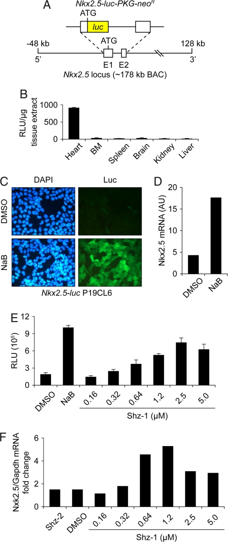
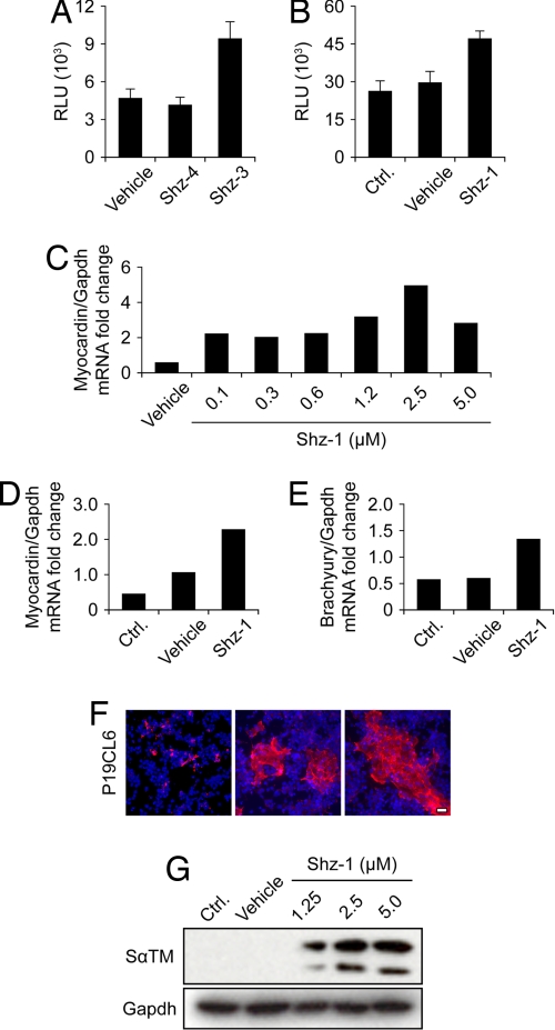
The clinical success of stem cell therapy for myocardial repair hinges on a better understanding of cardiac fate mechanisms. We have identified small molecules involved in cardiac fate by screening a chemical library for activators of the signature gene Nkx2.5, using a luciferase knockin bacterial artificial chromosome (BAC) in mouse P19CL6 pluripotent stem cells. We describe a family of sulfonyl-hydrazone (Shz) small molecules that can trigger cardiac mRNA and protein expression in a variety of embryonic and adult stem/progenitor cells, including human mobilized peripheral blood mononuclear cells (M-PBMCs). Small-molecule-enhanced M-PBMCs engrafted into the rat heart in proximity to an experimental injury improved cardiac function better than control cells. Recovery of cardiac function correlated with persistence of viable human cells, expressing human-specific cardiac mRNAs and proteins. Shz small molecules are promising starting points for drugs to promote myocardial repair/regeneration by activating cardiac differentiation in M-PBMCs.


Conflict of interest statement

The authors declare no conflict of interest.

Figures

Fig. 1.
Fig. 1.
Nkx2.5 cardiogenic small-molecule HTS in P19CL6 cells. (A) Schematic of reporter transgene with luciferase inserted by homologous recombination into the Nkx2.5 locus on an ≈178-kb mouse BAC. (B) In vitro luciferase assay of Nkx2.5-luc BAC transgenic mouse confirming cardiac tissue-restricted expression. (C) Nkx2.5-luc BAC transgenic P19CL6 cell line, clone #5-1, with specific induction of luciferase protein (detected by immunostaining) after exposure to 2.5 mM NaB for 3 days (Lower Right), compared with DMSO vehicle control, which showed a lack of luciferase staining (Upper Right). (D) NaB-mediated induction of Nkx2.5 mRNA by real-time RT-PCR in parental P19CL6 cells. (E) Dose-responsive activation of Nkx2.5-luc BAC by Shz-1 in clone #5-1 P19CL6 reporter cells at 48 h after drug exposure. Each data point represents the average + SD of 12 wells from a 96-well plate. (F) Dose-responsive activation of Nkx2.5 mRNA by Shz-1 in P19CL6 cells at 48 h after drug exposure, measured by real-time RT-PCR and normalized to gapdh mRNA levels.
Fig. 2.
Fig. 2.
Shz activation of myocardin reporter genes in P19CL6 and mouse ES cells. (A) Activation of myocardin-luc BAC by Shz-3 but not Shz-4 at 2.5 μM in transgenic P19CL6 reporter cells at 48 h after drug exposure. Each data point represents the average + SD of 12 wells from a 96-well plate. (B) Activation of endogenous myocardin tagged by homologous recombination with luciferase in SM1 mouse ES cell genome by Shz-1. Luciferase activity was measured 48 h after removal of LIF and exposure to Shz-1 at 2.5 μM. Each data point represents the average + SD of 12 wells from a 96-well plate. (C) Dose-responsive activation of myocardin mRNA by Shz-1 in P19CL6 cells at 48 h after drug exposure, measured by real-time RT-PCR and normalized to gapdh mRNA levels. (D) Activation of endogenous myocardin mRNA in SM1 ES cells by Shz-1 at 2.5 μM at 48 h after drug exposure and removal of LIF, measured by real time RT-PCR and normalized to GAPDH mRNA levels. (E) Activation of endogenous brachyury-T mRNA in SM1 ES cells by Shz-1 at 2.5 μM at 48 h after drug exposure and removal of LIF, measured by real-time RT-PCR and normalized to gapdh mRNA levels. (F) Expression of sarcomeric α-tropomyosin detected by CH1 mAb in vehicle (Left) or Shz-1 treated (Center and Right) P19CL6 cells at 2.5 μM after 72 h of drug exposure. (Scale bar, 30 μm.) (G) Dose-responsive activation of SαTM in P19CL6 cells by protein blot with CH1 mAb; GAPDH protein levels are shown to document equal loading.
Fig. 3.
Fig. 3.
Comparison of Shz and sodium butyrate and interrogation of known cardiogenic signaling pathways. (A) NaBut induces trimethylation of K27 of histone protein H3 and acetylation of total histone protein H4 but does not induce Sα-TM expression; the converse is true for Shz-1 in P19CL6 cells. (B) Like Shz-1, BMP-2 (50 ng/ml) activated the Nkx2.5-luc BAC in P19CL6 cells. However, the BMP-antagonist Noggin (500 ng/ml) did not significantly block Shz-1-mediated signaling at 48 h. Each data point is the average + SD of 12 wells from a 96-well plate. (C) Representative cell staining demonstrating induction of SαTM by Shz-1 (2.5 μM) (Left), GATA-4 by BMP-2 (50 ng/ml) (Center), and Oct3/4 by FGF-2 (20 ng/ml) (Right) after exposure to agent for 48 h.
Fig. 4.
Fig. 4.
Small-molecule treatment of human M-PBMCs in vitro. (A) Schematic of human M-PBMCs treatment protocol, 3 days with drug (10 μM) or vehicle control and then 7 days in drug-free media with media changes every 2–3 days. (B) Dose-responsive increase in M-PBMCs attachment/survival with increasing concentration of Shz-1 for the first 3 days or vehicle control; average cell counts on day 10 of at least 10 representative low-power fields (lpf) + SD (C) Activation of cardiac differentiation by Shz small molecules in human M-PBMCs in vitro. M-PBMCs were treated with vehicle or Shz-3 for 3 days, followed by 7 days in drug-free media; the cells were harvested and analyzed by RT-PCR for Nkx2.5, ANP, and gapdh transcripts; adult human heart mRNA was used a positive control. (D) M-PBMCs from a different donor were harvested before treatment (day 0) or treated with Shz-1 for 3 days, followed by 7 days in drug-free media, and then harvested and analyzed with heart muscle control by RT-PCR for cTnI and gapdh transcripts.
Fig. 5.
Fig. 5.
Functional rescue of cryoinjured rat heart by Shz-treated human M-PBMC xenografts. (A) Viable human M-PBMCs pretreated for 3 days with compound Shz-3 (at 5 μM) or vehicle control were injected by needle into the healthy myocardial perimeter of liquid nitrogen probe-mediated transmural burn injury, followed by 7 days in drug-free media. (B) Serial echocardiography was done at baseline (preinjury) and on days 3, 7, 14, and 21 after injury/xenografting, and the fractional shortening was calculated for each animal (n = 4, each group) and was compared with hearts that had received mock injection with media alone (no cells). At days 7, 14, and 21, the difference between Shz-3 small-molecule and vehicle-treated human M-PBMC xenografts was statistically significant, P = 0.00183, P = 0.00023, and P = 0.000238, respectively. (C) IHC of chimeric juxta-burn myocardial tissue from rat injected with in vitro DAPI-stained vehicle (Upper) or Shz-3-treated (Lower) human M-PBMCs by using a mAb that detects α-actinin exclusively in host rat myocardium (human cells evident by DAPI stained nuclei are negative for α-actinin) (Left) versus a human-specific cTnI mAb that exclusively detects viable human drug-induced (cardiac gene-expressing) cells in the needle-track (Lower Right). (Scale bar, 25 μm.) (Inset) Human M-PBMCs treated with drug and immunostained in vitro appear morphologically very similar to their in vivo counterparts. (D) RT-PCR of RNA from chimeric juxta-burn tissue of hearts injected with Shz-3 or vehicle-treated cells under high-stringency conditions that favor amplification of human versus rat Nkx2.5 or cTnI sequences. The control for RNA loading is 18S ribosomal RNA. Human and rat hearts are used as positive and negative controls, respectively.

References

    1. Sasaki K, et al. Ex vivo pretreatment of bone marrow mononuclear cells with endothelial NO synthase enhancer AVE9488 enhances their functional activity for cell therapy. Proc Natl Acad Sci USA. 2006;103:14537–14541. - PMC - PubMed
    1. Dimmeler S, et al. HMG-CoA reductase inhibitors (statins) increase endothelial progenitor cells via the PI 3-kinase/Akt pathway. J Clin Invest. 2001;108:391–397. - PMC - PubMed
    1. Wojakowski W, Kucia M, Kazmierski M, Ratajczak MZ, Tendera M. Circulating stem/progenitor cells in stable ischemic heart disease and acute coronary syndromes—relevant reparatory mechanism? Heart. 2008;94:27–33. - PubMed
    1. Rosenzweig A. Cardiac cell therapy—mixed results from mixed cells. New Engl J Med. 2006;355:1274–1277. - PubMed
    1. Assmus B, et al. Transcoronary transplantation of progenitor cells after myocardial infarction. New Engl J Med. 2006;355:1222–1232. - PubMed

Publication types

MeSH terms