. 2008 Jun;26(6):1646-55.
doi: 10.1634/stemcells.2007-0715. Epub 2008 Apr 17.

Transplantation of bone marrow-derived very small embryonic-like stem cells attenuates left ventricular dysfunction and remodeling after myocardial infarction

Affiliations

Transplantation of bone marrow-derived very small embryonic-like stem cells attenuates left ventricular dysfunction and remodeling after myocardial infarction

Buddhadeb Dawn et al. Stem Cells. 2008 Jun.

Abstract

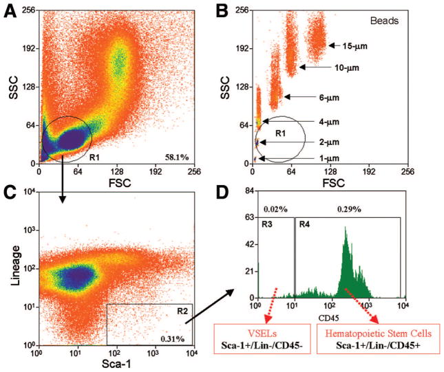
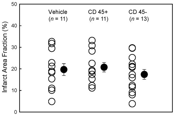
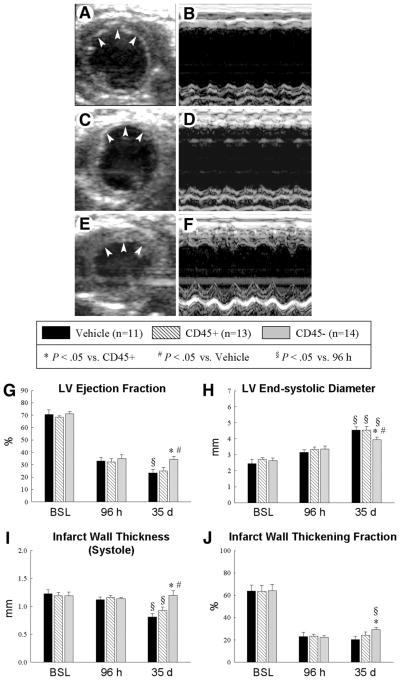
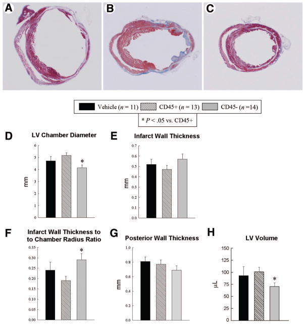
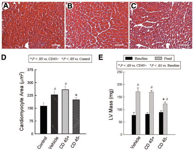
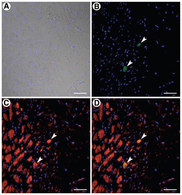
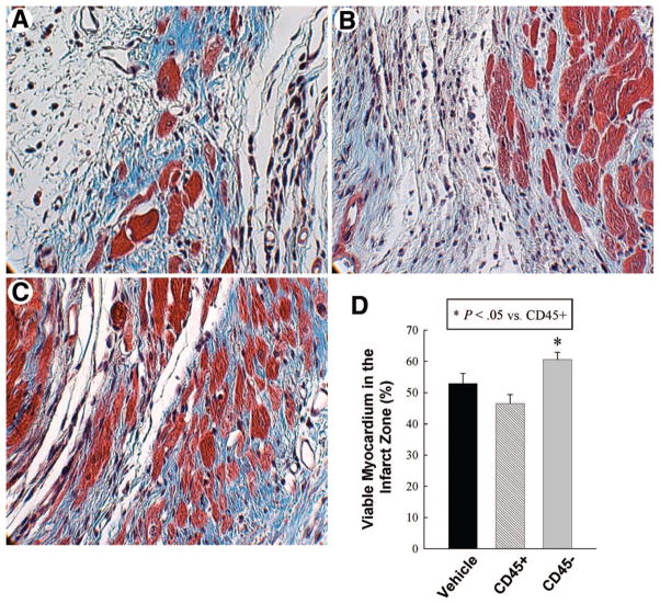
Adult bone marrow (BM) contains Sca-1+/Lin-/CD45- very small embryonic-like stem cells (VSELs) that express markers of several lineages, including cardiac markers, and differentiate into cardiomyocytes in vitro. We examined whether BM-derived VSELs promote myocardial repair after a reperfused myocardial infarction (MI). Mice underwent a 30-minute coronary occlusion followed by reperfusion and received intramyocardial injection of vehicle (n= 11), 1 x 10(5) Sca-1+/Lin-/CD45+ enhanced green fluorescent protein (EGFP)-labeled hematopoietic stem cells (n= 13 [cell control group]), or 1 x 10(4) Sca-1+/Lin-/CD45- EGFP-labeled cells (n= 14 [VSEL-treated group]) at 48 hours after MI. At 35 days after MI, VSEL-treated mice exhibited improved global and regional left ventricular (LV) systolic function (echocardiography) and attenuated myocyte hypertrophy in surviving tissue (histology and echocardiography) compared with vehicle-treated controls. In contrast, transplantation of Sca-1+/Lin-/CD45+ cells failed to confer any functional or structural benefits. Scattered EGFP+ myocytes and capillaries were present in the infarct region in VSEL-treated mice, but their numbers were very small. These results indicate that transplantation of a relatively small number of CD45- VSELs is sufficient to improve LV function and alleviate myocyte hypertrophy after MI, supporting the potential therapeutic utility of these cells for cardiac repair. Disclosure of potential conflicts of interest is found at the end of this article.


Conflict of interest statement

Disclosure of Potential Conflicts of Interest

The authors indicate no potential conflicts of interest.

Figures

Figure 1
Figure 1
Flow cytometric isolation of bone marrow-derived Sca-1+/Lin−/CD45+ hematopoietic stem cells and Sca-1+/Lin−/CD45− VSELs. Representative dot plots show sorting of small cells from the lymphoid gate (A) based on expression of Sca-1 (fluorescein isothiocyanate) and lineage markers (phycoerythrin) (C) and CD45 (APC-Cy7). (D): R3 contains Sca-1+/lin−/CD45− VSELs, whereas R4 contains Sca-1+/lin−/CD45+ cells. By comparing the sorting of bone marrow cells (BMCs) with the sorting of beads with known diameter, the FSC axis in (B) confirms the very small size (2–10 μm) of the cells in the region of interest in (A). As shown here (R3), only 0.02% of total BMCs are VSELs. Abbreviations: FSC, forward scatter characteristics; R, region; SSC, side scatter characteristics; VSEL, very small embryonic-like stem cell.
Figure 2
Figure 2
Myocardial infarct size. Myocardial infarct area fraction ([infarct area/left ventricular area] × 100) assessed from Masson’s trichrome-stained hearts in groups I–III, which were treated with vehicle, CD45+ hematopoietic stem cells, and very small embryonic-like stem cells, respectively. ○, individual mice; ●, mean ± SEM.
Figure 3
Figure 3
Echocardiographic assessment of LV function. Representative two-dimensional (A, C, E) and M-mode (B, D, F) images from vehicle-treated (A, B), CD45+ cell-treated (C, D), and very small embryonic-like stem cell (VSEL)-treated (E, F) mice 35 d after coronary occlusion/reperfusion. The infarct wall is delineated by arrowheads (A, C, E). Compared with the vehicle-treated and CD45+ cell-treated hearts, the VSEL-treated heart exhibited a smaller LV cavity, a thicker infarct wall, and improved motion of the infarct wall. Panels (G–J) demonstrate that transplantation of VSEL improved echocardiographic measurements of LV systolic function 35 d after myocardial infarction. Data are mean ± SEM. n = 11–14 mice per group. *, p < .05 versus group II at 35 d; #, p < .05 versus group I at 35 d; §, p < .05 versus values at 96 h in respective groups. Abbreviations: BSL, baseline; d, days; h, hours; LV, left ventricular.
Figure 4
Figure 4
Morphometric assessment of LV remodeling. Representative Masson’s trichrome-stained myocardial sections from vehicle-treated (A), CD45+ hematopoietic stem cell-treated (B), and very small embryonic-like stem cell (VSEL)-treated (C) hearts. Scar tissue and viable myocardium are identified in blue and red, respectively. Note that the LV cavity is smaller and the infarct wall thicker in the VSEL-treated heart. Panels (D–H) illustrate morphometric measurements of LV structural parameters. Data are mean ± SEM. n = 11–14 mice per group. *, p < .05 versus group II. Abbreviation: LV, left ventricular.
Figure 5
Figure 5
Assessment of cardiomyocyte and LV hypertrophy. Panels (A–C) show representative images of cardiomyocytes in the viable myocardium from Masson’s trichrome-stained vehicle-treated (A), CD45+ hematopoietic stem cell-treated (B), and very small embryonic-like stem cell (VSEL)-treated hearts (C). Scale bars = 50 μm. In contrast to CD45+ hematopoietic stem cell-treated hearts, VSEL-treated hearts did not exhibit increased myocyte cross-sectional area compared with noninfarcted control hearts (D). Echocardiographically estimated LV mass was significantly less in VSEL-treated hearts (E). Data are mean ± SEM. n = 11–14 mice per group. (D): *, p < .05 versus group II; #, p < .05 versus control; (E): *, p < .05 versus group II and III (final); #, p < .05 versus respective baseline values. Abbreviation: LV, left ventricular.
Figure 6
Figure 6
Very small embryonic-like stem cell (VSEL) transplantation and cardiomyocyte regeneration. VSELs and myocytes are identified by enhanced green fluorescent protein (EGFP) ([B, D], green) and α-sarcomeric actin ([C, D], red), respectively; (D) shows the merged image. Two myocytes are shown that are positive for both EGFP (arrowheads; [B], green) and α-sarcomeric actin (arrowheads; [C], red). Nuclei were stained with 4,6-diamidino-2-phenylindole ([A, D], blue). Scale bars = 40 μm.
Figure 7
Figure 7
Assessment of myocyte area fraction in the infarct area. Panels (A–C) illustrate representative examples of scar in Masson’s trichrome-stained, vehicle-treated (A), CD45+ hematopoietic stem cell-treated (B), and very small embryonic-like stem cell-treated (C) hearts. Magnification, ×600. Quantitative data are presented in (D). Data are mean ± SEM. n = 11–14 mice per group. *, p < .05 versus group II.

References

    1. Bolli R, Jneid H, Dawn B. Bone marrow cell-mediated cardiac regeneration a veritable revolution. J Am Coll Cardiol. 2005;46:1659–1661. - PubMed
    1. Abdel-latif A, Bolli R, Tleyjeh IM, et al. Adult bone marrow-derived cells for cardiac repair: A systematic review and meta-analysis. Arch Int Med. 2007;167:989–997. - PubMed
    1. Min JY, Yang Y, Converso KL, et al. Transplantation of embryonic stem cells improves cardiac function in postinfarcted rats. J Appl Physiol. 2002;92:288–296. - PubMed
    1. Kofidis T, de Bruin JL, Yamane T, et al. Insulin-like growth factor promotes engraftment, differentiation, and functional improvement after transfer of embryonic stem cells for myocardial restoration. Stem Cells. 2004;22:1239–1245. - PubMed
    1. Leor J, Gerecht S, Cohen S, et al. Human embryonic stem cell transplantation to repair the infarcted myocardium. Heart. 2007;93:1278–1284. - PMC - PubMed

Publication types

MeSH terms

Substances