. 2008 Apr 18;320(5874):365-9.
doi: 10.1126/science.1151250.

Wnt5a control of cell polarity and directional movement by polarized redistribution of adhesion receptors

Affiliations

Wnt5a control of cell polarity and directional movement by polarized redistribution of adhesion receptors

Eric S Witze et al. Science. .

Abstract

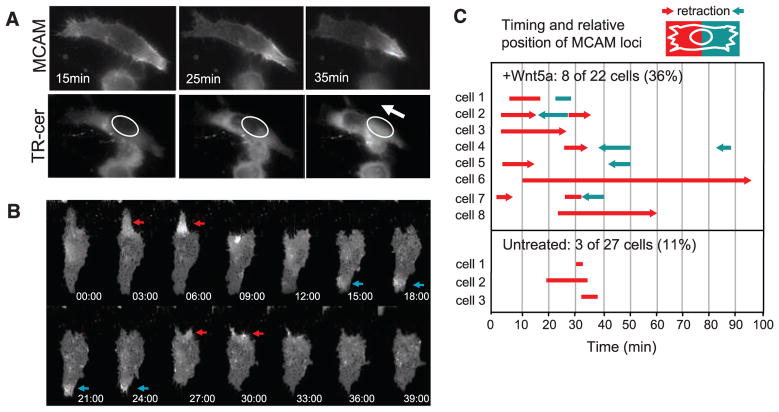
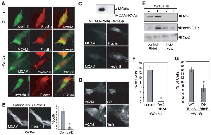
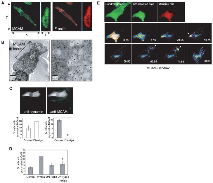
Mechanisms by which Wnt pathways integrate the organization of receptors, organelles, and cytoskeletal proteins to confer cell polarity and directional cell movement are incompletely understood. We show that acute responses to Wnt5a involve recruitment of actin, myosin IIB, Frizzled 3, and melanoma cell adhesion molecule into an intracellular structure in a melanoma cell line. In the presence of a chemokine gradient, this Wnt-mediated receptor-actin-myosin polarity (W-RAMP) structure accumulates asymmetrically at the cell periphery, where it triggers membrane contractility and nuclear movement in the direction of membrane retraction. The process requires endosome trafficking, is associated with multivesicular bodies, and is regulated by Wnt5a through the small guanosine triphosphatases Rab4 and RhoB. Thus, cell-autonomous mechanisms allow Wnt5a to control cell orientation, polarity, and directional movement in response to positional cues from chemokine gradients.


Figures

Fig. 1
Fig. 1
An asymmetric “W-RAMP structure” forms in response to Wnt5a and defines cell polarization and orientation. (A) Examples of cells with uniform MCAM localization versus asymmetric W-RAMP structures, observed by indirect immunofluorescence in WM239A cells treated for 30 min with or without Wnt5a or in WM1789 cells treated for 30 min with Wnt5a. Nuclei are shown by 4′,6′-diamidino-2-phenylindole (DAPI) staining. (B) Wnt5a significantly increases the number of WM239A cells displaying asymmetric MCAM compared with control or Wnt3a-treated cells. [Data show means ± SEM. *P < 0.005 by Student’s t test. Number of experiments (n) = 4. Cells counted: control, 1391; Wnt5a, 1380; Wnt3a, 1408.] RNAi knockdown of endogenous Wnt5a reduces the number of cells exhibiting the W-RAMP structure under untreated conditions, from 6% to less than 0.9% (Data show means with highest and lowest values, n = 2. Cells counted: control, 648; Wnt5a-RNAi, 1051). (C) In the presence of CXCL12, Wnt5a increased the percentage of cells with W-RAMP structures (red) positioned distal to Golgi (green) (means ± SEM; *P < 0.005, n = 4. Cells counted: control, 99; Wnt5a, 136). In the absence of CXCL12, Wnt5a did not affect the W-RAMP structure position relative to Golgi (n = 3. Cells counted: control, 134; Wnt5a, 131). (D) Wnt5a increased the number of cells with Golgi pointed toward or away from the gradient (class 1 and 3), with little directional bias.
Fig. 2
Fig. 2
Formation of the W-RAMP structure is dynamic and associated with membrane retraction. (A) Live WM239A cells were incubated with AlexaFluor-488–conjugated MCAM antibody and Texas Red (TR)–ceramide. In response to Wnt5a (in absence of CXCL12) MCAM decreases at the left end of the cell and increases at the right, followed by membrane retraction toward the W-RAMP structure. TR-ceramide staining reveals nuclear translocation in the direction of membrane retraction (arrow). (B) The W-RAMP structure forms in successive waves. MCAM-GFP expressed in WM239A cells accumulates at one cell end (red arrow) followed by membrane retraction (after 9 min). A second W-RAMP structure forms at the opposite cell end (blue arrow), followed by membrane retraction (after 27 min). A third W-RAMP structure (red arrow) is followed by membrane retraction at 30 min. (C) Live cells were monitored continuously for MCAM-GFP localization. The x axis represents time after addition of Wnt5a. The length of each bar represents the duration of each wave of polarized accumulation of MCAM. Red versus blue lines distinguish the relative end of the cell at which the wave forms. Arrowheads indicate cases where membrane retraction follows formation of a wave, and the relative end of the cell at which the retraction occurs. Representative cells can be viewed in movies S7 to S12.
Fig. 3
Fig. 3
The W-RAMP structure is formed through reciprocal interactions with actin and myosin IIB. (A) In Wnt5a-treated cells, myosin IIB and F-actin form asymmetric structures that partially overlap with the W-RAMP structure. (B) Latrunculin B (10 nM) pretreatment of cells strongly inhibits Wnt5a-dependent formation of the W-RAMP structure (means ± SEM; *P < 0.005. Cells counted: dimethylsulfoxide-treated, 1362; LatB, 1538). (C) RNAi knockdown of MCAM expression leads to actin structures that are more diffuse and disorganized than controls, as well as complete abrogation of myosin IIB structures. (D) Indirect immunofluorescence of WM239A cells shows that Fz3 colocalizes with the W-RAMP structure, but transferrin receptor does not. (E) Pull-down assay with immobilized glutathione S-transferase–rhotekin reveals elevated RhoB-GTP in response to Wnt5a. Dvl2 knockdown blocks the activation of RhoB by Wnt5a. (F) Dvl2 RNAi knockdown abolishes W-RAMP structure in Wnt5a-treated cells (*P < 0.05, n = 3. Cells counted: control RNAi, 1002; Dvl2-RNAi, 1139). (G) Transient transfection of cells with DN-RhoB blocks Wnt5a-dependent formation of the W-RAMP structure (*P < 0.05, n = 3. Cells counted: WT-RhoB, 608; DN-RhoB, 590).
Fig. 4
Fig. 4
Formation and dynamic movement of the W-RAMP structure requires membrane internalization and endosome recycling and is associated with multivesicular bodies. (A) Confocal Z-series reconstructed in the region of the MCAM structure (blue line), showed that MCAM (MCAM-specific antibody, green) and F-actin (phalloidin, red) are localized to both cytosolic and membrane regions and are excluded from the nucleus. (B) Electron micrographs show accumulation of membrane vesicles associated with the W-RAMP structure. (Right) Higher magnification reveals that vesicles consist of multivesicular bodies (asterisks) interspersed among cytoskeletal filaments. (C) Cells expressing DN-dynamin, easily distinguished by their higher immunoreactivity, showed higher percentages with uniform symmetric MCAM localization and lower percentages with asymmetric MCAM localization (*P < 0.005, n = 3. Cells counted: control, 167; DN-dynamin, 203). (D) DN-Rab4 blocks the ability of Wnt5a to promote formation of W-RAMP structures, decreased significantly compared with Wnt5a alone (*P < 0.05, n = 4. Cells counted: control, 1019; Wnt5a, 1244; DN-Rab4, 1262; DN-Rab4+Wnt5a, 1034). (E) Red fluorescence from photoactivated MCAM-Dendra2 accumulates transiently in a perinuclear pool by 60 min, then at the opposite end of the cell by 71 min (arrow), followed by membrane retraction. Colors from lowest to highest intensity are black, blue, green, yellow, orange, red, and white.

Comment in

References

    1. Adler PN. Dev Cell. 2002;2:525. - PubMed
    1. Shimada Y, Yonemura S, Ohkura H, Strutt D, Uemura T. Dev Cell. 2006;10:209. - PubMed
    1. Park FD, Tenlen JR, Priess JR. Curr Biol. 2004;14:2252. - PubMed
    1. Goldstein B, Takeshita H, Mizumoto K, Sawa H. Dev Cell. 2006;10:391. - PMC - PubMed
    1. Hilliard MA, Bargmann CI. Dev Cell. 2006;10:379. - PubMed

Publication types

MeSH terms