Structural basis of toll-like receptor 3 signaling with double-stranded RNA
- PMID: 18420935
- PMCID: PMC2761030
- DOI: 10.1126/science.1155406
Structural basis of toll-like receptor 3 signaling with double-stranded RNA
Abstract
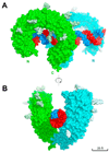
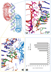
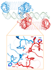
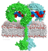
Toll-like receptor 3 (TLR3) recognizes double-stranded RNA (dsRNA), a molecular signature of most viruses, and triggers inflammatory responses that prevent viral spread. TLR3 ectodomains (ECDs) dimerize on oligonucleotides of at least 40 to 50 base pairs in length, the minimal length required for signal transduction. To establish the molecular basis for ligand binding and signaling, we determined the crystal structure of a complex between two mouse TLR3-ECDs and dsRNA at 3.4 angstrom resolution. Each TLR3-ECD binds dsRNA at two sites located at opposite ends of the TLR3 horseshoe, and an intermolecular contact between the two TLR3-ECD C-terminal domains coordinates and stabilizes the dimer. This juxtaposition could mediate downstream signaling by dimerizing the cytoplasmic Toll interleukin-1 receptor (TIR) domains. The overall shape of the TLR3-ECD does not change upon binding to dsRNA.
Figures


Similar articles
-
The toll-like receptor 3:dsRNA signaling complex.Biochim Biophys Acta. 2009 Sep-Oct;1789(9-10):667-74. doi: 10.1016/j.bbagrm.2009.06.005. Epub 2009 Jul 9. Biochim Biophys Acta. 2009. PMID: 19595807 Free PMC article. Review.
-
The dsRNA binding site of human Toll-like receptor 3.Proc Natl Acad Sci U S A. 2006 Jun 6;103(23):8792-7. doi: 10.1073/pnas.0603245103. Epub 2006 May 23. Proc Natl Acad Sci U S A. 2006. PMID: 16720699 Free PMC article.
-
A second binding site for double-stranded RNA in TLR3 and consequences for interferon activation.Nat Struct Mol Biol. 2008 Jul;15(7):761-3. doi: 10.1038/nsmb.1453. Epub 2008 Jun 22. Nat Struct Mol Biol. 2008. PMID: 18568036
-
Insights into the dynamic nature of the dsRNA-bound TLR3 complex.Sci Rep. 2019 Mar 6;9(1):3652. doi: 10.1038/s41598-019-39984-8. Sci Rep. 2019. PMID: 30842554 Free PMC article.
-
Beyond dsRNA: Toll-like receptor 3 signalling in RNA-induced immune responses.Biochem J. 2014 Mar 1;458(2):195-201. doi: 10.1042/BJ20131492. Biochem J. 2014. PMID: 24524192 Review.
Cited by
-
Targeting the innate immune receptor TLR8 using small-molecule agents.Acta Crystallogr D Struct Biol. 2020 Jul 1;76(Pt 7):621-629. doi: 10.1107/S2059798320006518. Epub 2020 Jun 17. Acta Crystallogr D Struct Biol. 2020. PMID: 32627735 Free PMC article. Review.
-
AMD Genetics in India: The Missing Links.Front Aging Neurosci. 2016 May 23;8:115. doi: 10.3389/fnagi.2016.00115. eCollection 2016. Front Aging Neurosci. 2016. PMID: 27252648 Free PMC article. Review.
-
Structure and Emerging Functions of LRCH Proteins in Leukocyte Biology.Front Cell Dev Biol. 2020 Sep 22;8:584134. doi: 10.3389/fcell.2020.584134. eCollection 2020. Front Cell Dev Biol. 2020. PMID: 33072765 Free PMC article. Review.
-
Dissecting novel mechanisms of hepatitis B virus related hepatocellular carcinoma using meta-analysis of public data.World J Gastrointest Oncol. 2022 Sep 15;14(9):1856-1873. doi: 10.4251/wjgo.v14.i9.1856. World J Gastrointest Oncol. 2022. PMID: 36187396 Free PMC article.
-
Leucine-rich repeat 11 of Toll-like receptor 9 can tightly bind to CpG-containing oligodeoxynucleotides, and the positively charged residues are critical for the high affinity.J Biol Chem. 2012 Aug 31;287(36):30596-609. doi: 10.1074/jbc.M112.396432. Epub 2012 Jul 20. J Biol Chem. 2012. PMID: 22822061 Free PMC article.
References
-
- Kaisho T, Akira S. J. Allergy Clin. Immunol. 2006;117:979. - PubMed
-
- Iwasaki A, Medzhitov R. Nat. Immunol. 2004;5:987. - PubMed
-
- Gay NJ, Gangloff M. Annu. Rev. Biochem. 2007;76:141. - PubMed
-
- Alexopoulou L, Holt AC, Medzhitov R, Flavell RA. Nature. 2001;413:732. - PubMed
-
- Oshiumi H, Matsumoto M, Funami K, Akazawa T, Seya T. Nat. Immunol. 2003;4:161. - PubMed
Publication types
MeSH terms
Substances
Associated data
- Actions
- Actions
Grants and funding
LinkOut - more resources
Full Text Sources
Other Literature Sources
Molecular Biology Databases

