. 2008 Jul 15;178(2):158-67.
doi: 10.1164/rccm.200711-1661OC. Epub 2008 Apr 17.

Surfactant proteins A and D suppress alveolar macrophage phagocytosis via interaction with SIRP alpha

Affiliations

Surfactant proteins A and D suppress alveolar macrophage phagocytosis via interaction with SIRP alpha

William J Janssen et al. Am J Respir Crit Care Med. .

Abstract

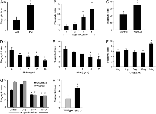
Rationale: Efficient removal of apoptotic cells is essential for the resolution of acute pulmonary inflammation. Alveolar macrophages ingest apoptotic cells less avidly than other professional phagocytes at rest but overcome this defect during acute inflammation. Surfactant protein (SP)-A and SP-D are potent modulators of macrophage function and may suppress clearance of apoptotic cells through activation of the transmembrane receptor signal inhibitory regulatory protein alpha (SIRP alpha).

Objectives: To investigate whether binding of SP-A and SP-D to SIRP alpha on alveolar macrophages suppresses apoptotic cell clearance.

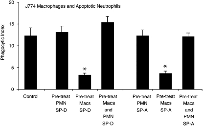
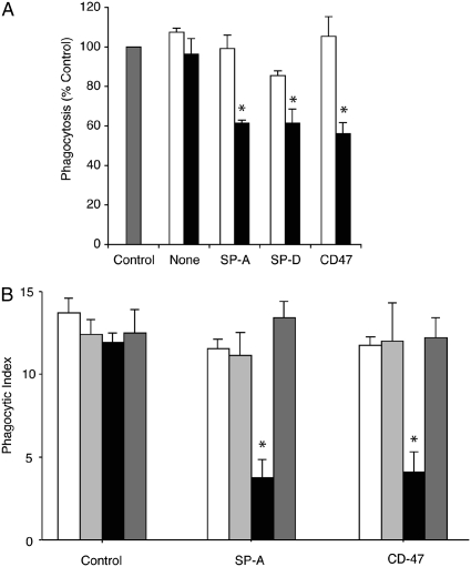
Methods: Phagocytosis of apoptotic cells was assessed using macrophages pretreated with SP-A, SP-D, or the collectin-like molecule C1q. Binding of SP-A and SP-D to SIRP alpha was confirmed in vitro using blocking antibodies and fibroblasts transfected with active and mutant SIRP alpha. The effects of downstream molecules SHP-1 and RhoA on phagocytosis were studied using SHP-1-deficient mice, sodium stibogluconate, and a Rho kinase inhibitor. Lipopolysaccharide was given to chimeric mice to study the effects of SP-A and SP-D binding on inflammatory macrophages.

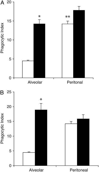
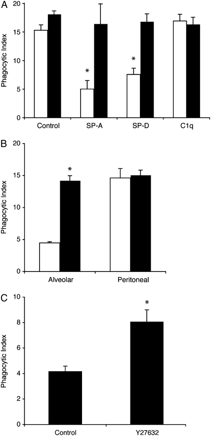
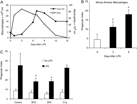
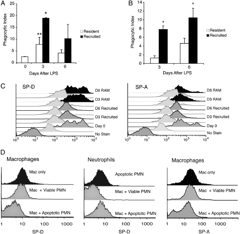
Measurements and main results: Preincubation of macrophages with SP-A or SP-D suppressed apoptotic cell clearance. Surfactant suppression of macrophage phagocytosis was reversed by blocking SIRP alpha and inhibiting downstream molecules SHP-1 and RhoA. Macrophages from inflamed lungs ingested apoptotic cells more efficiently than resting alveolar macrophages. Recruited mononuclear phagocytes with low levels of SP-A and SP-D mediated this effect.

Conclusions: SP-A and SP-D tonically inhibit alveolar macrophage phagocytosis by binding SIRP alpha. During acute pulmonary inflammation, defects in apoptotic cell clearance are overcome by recruited mononuclear phagocytes.


Figures

<b>Figure 1.</b>
Figure 1.
Lung collectins contribute to tonic suppression of alveolar macrophages (AMs). (A) Murine AMs and peritoneal macrophages (PMs) were isolated from Institute for Cancer Research mice and cocultured with apoptotic Jurkat T cells for 1 hour. *Significantly different from AMs (P < 0.05). (B) Murine AMs were cultured for the times indicated and then coincubated with apoptotic Jurkat T cells. **Significantly different from Day 1 phagocytic index (P < 0.05). (C) Human AMs were cultured directly (control) or after washing with phosphate-buffered saline (PBS). Washed and unwashed cells were incubated overnight. Phagocytosis was assessed after a 90-minute coculture with apoptotic human neutrophils. *Significantly different from unwashed AMs (P < 0.05). (DF) Dose–response curves for the lung collectins were created by adding surfactant protein (SP)-D, SP-A, and C1q to cultured J774 macrophages at the indicated concentrations for 20 minutes. The cells were gently washed to remove unbound protein, and then fresh medium containing apoptotic Jurkat T cells was added. Phagocytosis was assessed 90 minutes later. *P < 0.05 versus control. (G) Human AMs were cultured directly after isolation or after washing with PBS. SP-A, SP-D, or C1q (10 μg/ml) was added for 20 minutes, media were removed, and fresh media containing viable or apoptotic Jurkat T cells were added. Phagocytosis was significantly reduced by SP-A and SP-D for washed (*P < 0.05) and unwashed cells (**P < 0.05) versus control. (H) Mouse AMs were isolated from wild-type or SP-D–deficient mice. The macrophages were plated overnight and then fed apoptotic Jurkat T cells for 90 minutes. *P < 0.05.
<b>Figure 2.</b>
Figure 2.
Lung collectins serve dual regulatory roles in macrophage clearance of apoptotic cells. J774A.1 macrophages or apoptotic human neutrophils (PMN) were coincubated with SP-A or SP-D (10 μg/ml) for 20 minutes and then washed to remove excess, unbound collectins. Phagocytosis assays were performed after pairing: (1) collectin-naive macrophages + collectin-treated PMN, (2) collectin-treated macrophages + collectin-naive apoptotic PMN, or (3) collectin-treated macrophages + collectin-treated PMN. The control group is composed of collectin-naive macrophages and apoptotic neutrophils. Graphs represent the mean phagocytic index ± SEM of three independent experiments (*P < 0.05).
<b>Figure 3.</b>
Figure 3.
Signal inhibitory regulatory protein α (SIRPα) is required for collectin-mediated suppression of phagocytosis. (A) J774A.1 macrophages were incubated with a SIRPα blocking antibody or a rat IgG1 isotype control for 30 minutes before the addition of surfactant protein (SP)-A or SP-D (10 μg/ml). Excess media were removed and fresh media containing apoptotic Jurkat T cells were added for 90 minutes, then phagocytosis was assessed. Open bars, anti-SIRP antibody; solid bars, rat IgG isotype; shaded bar, control. *Significantly different from control (P < 0.05). (B) Swiss 3T3 fibroblasts were transiently transfected with an empty vector, an active SIRPα vector, or an inactive SIRPα construct. Forty-eight hours after transfection, culture media were changed and SP-A or CD47-Fc ligand (10 μg/ml) was added for 20 minutes. Cells were gently washed once and then apoptotic cells were added. Phagocytosis of apoptotic cells was only decreased in cells transfected with the active SIRPα construct and exposed to SP-A or CD47-Fc ligand (*P < 0.01). Results are expressed as the phagocytic index ± SEM for three separate experiments. Open bars, control; light shaded bars, empty vector; solid bars, SIRP; dark shaded bars, inactive SIRP.
<b>Figure 4.</b>
Figure 4.
Src homology protein (SHP) inhibition restores phagocytic capacity of alveolar macrophages (AMs). (A) Murine alveolar and peritoneal macrophages were isolated from ICR mice and cultured overnight. Sodium stibogluconate was added to inhibit SHP activity. Media were removed and fresh media containing apoptotic Jurkat T cells were added. Compared with peritoneal macrophages (PMs), AMs exhibited significantly reduced phagocytosis at baseline (**P < 0.001). This defect was corrected by SHP inhibition (*P < 0.05 vs. resting control). Open bars, control; solid bars, SHP inhibitor. (B) AMs and PMs were isolated from 4-week-old motheaten mice or age-matched C57 BL/6 controls. After overnight culture, phagocytosis of apoptotic Jurkat T cells was assessed (*P < 0.05 for wild-type AMs compared with viable motheaten AMs). Open bars, wild-type; solid bars, motheaten.
<b>Figure 5.</b>
Figure 5.
Surfactant proteins inhibit efferocytosis through activation of RhoA. (A) J774A.1 macrophages were cultured in the presence of the Rho kinase inhibitor Y27632 (10 μM for 20 min) and then treated with 10 μg/ml surfactant protein (SP)-A, SP-D, or C1q. Phagocytosis of apoptotic Jurkat T cells was assessed after 90 minutes of coincubation; *P < 0.05 versus control. (B) Alveolar macrophages and peritoneal macrophages were isolated from ICR mice, cultured in the presence of Y27632, and then fed apoptotic Jurkat T cells. (A, B) Open bars, control; solid bars, Y27632, (C) Mice were treated with 10 mg/kg Y27632 or phosphate-buffered saline by gavage. Four hours later, apoptotic murine thymocytes were instilled directly into the lungs. Bronchoalveolar lavage was performed 60 minutes later and the phagocytic index was assessed on cytospin samples using light microscopy. n = 8 mice per group. *P < 0.05 versus control.
<b>Figure 6.</b>
Figure 6.
Phagocytosis increases during inflammation. (A) Two hundred micrograms of LPS in 50 μl phosphate-buffered saline was administered to C57BL/6 mice by direct intratracheal instillation. Bronchoalveolar lavage (BAL) was performed at the times indicated. Absolute macrophage and neutrophil counts were obtained by multiplying BAL leukocyte counts by differential cell counts. Values represent the mean of three independent experiments with a minimum of four mice per group. (B) Macrophages from LPS-treated mice were purified from BAL by density centrifugation. After overnight culture, 5 × 10⁶ apoptotic Jurkat T cells were added to each well. Phagocytosis was assayed 90 minutes later. (C) Macrophages were isolated from uninjured mice (no LPS) or 6 days after treatment with intratracheal LPS. Purified alveolar macrophages (AMs) were cultured overnight, and then treated with surfactant protein (SP)-A, SP-D, or C1q (10 μg/ml) for 20 minutes. Phagocytosis of apoptotic Jurkat T cells was assessed. (*P < 0.05 versus control.) PMN = neutrophils.
<b>Figure 7.</b>
Figure 7.
Phagocytosis of apoptotic cells by resident and recruited macrophages during inflammation. LPS was administered to green fluorescent protein (GFP)–expressing chimeric mice. Resident alveolar macrophages (RAMs) and recruited mononuclear phagocytes were differentiated using flow cytometry based on GFP autofluorescence. (A) Resident and recruited AMs were fluorescently sorted, cultured overnight, and then coincubated with apoptotic Jurkat T cells. Phagocytosis was assessed 60 minutes later; *P < 0.05 for recruited versus resident macrophages; **P < 0.05 for inflammatory resident macrophages versus baseline. (B) Cytospin samples of freshly sorted resident and recruited macrophages were stained with Wright's Giemsa stain and analyzed by light microscopy for phagocytosis of endogenous apoptotic cells. The phagocytic index for this experiment represents macrophages that ingested apoptotic neutrophils in vivo; *P < 0.05 for recruited versus resident macrophages. (C) Flow cytometry was performed on lavage specimens from LPS-treated mice and controls (Day 0) using antibodies to surfactant protein (SP)-A and SP-D. RAMs (black) and recruited mononuclear phagocytes (gray) were identified by forward scatter, side scatter, and FL1 autofluorescence (GFP). Isotype controls are shown below each sample. Day 0 samples only contain RAMs. (D) Fluorescein isothiocyanate–labeled SP-D was added to J774 macrophages alone, apoptotic neutrophils alone, viable neutrophils alone, or to 1:1 mixtures containing neutrophils and macrophages for 30 minutes on ice. Samples were washed, fixed, and analyzed by flow cytometry. J774 macrophages were prelabeled with the fluorescent dye PKH26-PCL to permit discrimination between macrophages and neutrophils with flow cytometry. Experiments were repeated with SP-A. Histograms represent findings from three independent experiments. PMN = human neutrophils.

References

    1. Fadok VA, Bratton DL, Konowal A, Freed PW, Westcott JY, Henson PM. Macrophages that have ingested apoptotic cells in vitro inhibit proinflammatory cytokine production through autocrine/paracrine mechanisms involving TGF-beta, PGE2, and PAF. J Clin Invest 1998;101:890–898. - PMC - PubMed
    1. Morimoto K, Amano H, Sonoda F, Baba M, Senba M, Yoshimine H, Yamamoto H, Ii T, Oishi K, Nagatake T. Alveolar macrophages that phagocytose apoptotic neutrophils produce hepatocyte growth factor during bacterial pneumonia in mice. Am J Respir Cell Mol Biol 2001;24:608–615. - PubMed
    1. Golpon HA, Fadok VA, Taraseviciene-Stewart L, Scerbavicius R, Sauer C, Welte T, Henson PM, Voelkel NF. Life after corpse engulfment: phagocytosis of apoptotic cells leads to VEGF secretion and cell growth. FASEB J 2004;18:1716–1718. - PubMed
    1. Cox G, Crossley J, Xing Z. Macrophage engulfment of apoptotic neutrophils contributes to the resolution of acute pulmonary inflammation in vivo. Am J Respir Cell Mol Biol 1995;12:232–237. - PubMed
    1. Haslett C. Granulocyte apoptosis and its role in the resolution and control of lung inflammation. Am J Respir Crit Care Med 1999;160:S5–S11. - PubMed

Publication types

Substances