

Mechanical load and BMP signaling during tendon repair: a role for follistatin?
- PMID: 18421531
- PMCID: PMC2505240
- DOI: 10.1007/s11999-008-0253-0
Mechanical load and BMP signaling during tendon repair: a role for follistatin?
Abstract
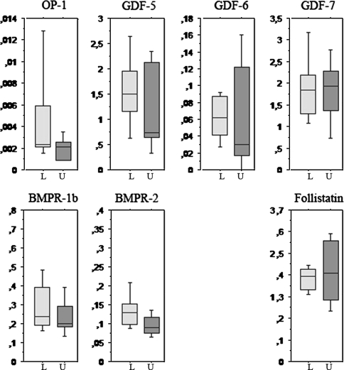
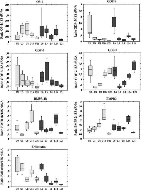
Healing of the rat Achilles tendon is sensitive to mechanical loading, and the callus strength is reduced by 3/4 after 14 days, if loading is prevented. Exogenous GDFs stimulate tendon healing. This response is influenced by loading: without loading, cartilage and bone formation is initiated. This implies BMP signaling is crucial during tendon healing and influenced by mechanical loading. We therefore asked if mechanical loading influences the gene expression of the BMP signaling system in intact and healing tendons, and how the BMP signaling system changes during healing. The genes were four BMPs (OP-1/BMP-7, GDF-5/CDMP-1/BMP-14, GDF-6/CDMP-2/BMP-13, and GDF-7/CDMP-3/BMP-12), two receptors (BMPR1b and BMPR2), and the antagonists follistatin and noggin. The Achilles tendon was transected in rats and left to heal. Half of the rats had one Achilles tendon unloaded by injection of Botox in the calf muscles. Ten tendons were analyzed before transection and for each of four time points. All genes except noggin were expressed at all time points, but followed different patterns during healing. Loading strongly decreased the expression of follistatin, which could lead to increased signaling. The BMP system appears involved in tendon maintenance and healing, and may respond to mechanical loading.
Figures
References
-
- {'text': '', 'ref_index': 1, 'ids': [{'type': 'PubMed', 'value': '10191749', 'is_inner': True, 'url': 'https://pubmed.ncbi.nlm.nih.gov/10191749/'}]}
- Aspenberg P, Forslund C. Enhanced tendon healing with GDF 5 and 6. Acta Orthop Scand. 1999;70:51–54. - PubMed
-
- {'text': '', 'ref_index': 1, 'ids': [{'type': 'DOI', 'value': '10.1016/S0736-0266(03)00049-4', 'is_inner': False, 'url': 'https://doi.org/10.1016/s0736-0266(03)00049-4'}, {'type': 'PubMed', 'value': '12919870', 'is_inner': True, 'url': 'https://pubmed.ncbi.nlm.nih.gov/12919870/'}]}
- Chhabra A, Tsou D, Clark RT, Gaschen V, Hunziker EB, Mikic B. GDF-5 deficiency in mice delays Achilles tendon healing. J Orthop Res. 2003;21:826–835. - PubMed
-
- {'text': '', 'ref_index': 1, 'ids': [{'type': 'DOI', 'value': '10.3109/03008200109005648', 'is_inner': False, 'url': 'https://doi.org/10.3109/03008200109005648'}, {'type': 'PubMed', 'value': '11913489', 'is_inner': True, 'url': 'https://pubmed.ncbi.nlm.nih.gov/11913489/'}]}
- Clark RT, Johnson TL, Schalet BJ, Davis L, Gaschen V, Hunziker EB, Oldberg A, Mikic B. GDF-5 deficiency in mice leads to disruption of tail tendon form and function. Connect Tissue Res. 2001;42:175–186. - PubMed
-
- {'text': '', 'ref_index': 1, 'ids': [{'type': 'DOI', 'value': '10.1007/s004290050286', 'is_inner': False, 'url': 'https://doi.org/10.1007/s004290050286'}, {'type': 'PubMed', 'value': '10460474', 'is_inner': True, 'url': 'https://pubmed.ncbi.nlm.nih.gov/10460474/'}]}
- D’Souza D, Patel K. Involvement of long- and short-range signalling during early tendon development. Anat Embryol (Berl). 1999;200:367–375. - PubMed
-
- {'text': '', 'ref_index': 1, 'ids': [{'type': 'DOI', 'value': '10.1152/japplphysiol.01333.2006', 'is_inner': False, 'url': 'https://doi.org/10.1152/japplphysiol.01333.2006'}, {'type': 'PubMed', 'value': '17412787', 'is_inner': True, 'url': 'https://pubmed.ncbi.nlm.nih.gov/17412787/'}]}
- Eliasson P, Fahlgren A, Pasternak B, Aspenberg P. Unloaded rat Achilles tendons continue to grow, but lose viscoelasticity. J Appl Physiol. 2007;103:459–463. - PubMed
MeSH terms
Substances
LinkOut - more resources
Full Text Sources
Other Literature Sources
Miscellaneous
