. 2008 Apr 19:8:114.
doi: 10.1186/1471-2148-8-114.

Diversification and adaptive sequence evolution of Caenorhabditis lysozymes (Nematoda: Rhabditidae)

Affiliations

Diversification and adaptive sequence evolution of Caenorhabditis lysozymes (Nematoda: Rhabditidae)

Hinrich Schulenburg et al. BMC Evol Biol. .

Abstract

Background: Lysozymes are important model enzymes in biomedical research with a ubiquitous taxonomic distribution ranging from phages up to plants and animals. Their main function appears to be defence against pathogens, although some of them have also been implicated in digestion. Whereas most organisms have only few lysozyme genes, nematodes of the genus Caenorhabditis possess a surprisingly large repertoire of up to 15 genes.

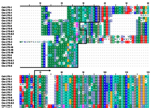
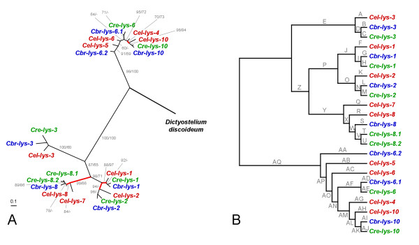
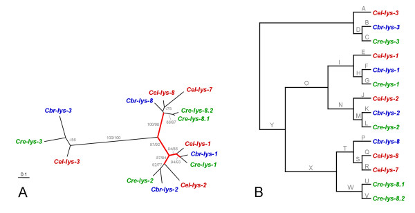
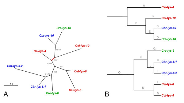
Results: We used phylogenetic inference and sequence analysis tools to assess the evolution of lysozymes from three congeneric nematode species, Caenorhabditis elegans, C. briggsae, and C. remanei. Their lysozymes fall into three distinct clades, one belonging to the invertebrate-type and the other two to the protist-type lysozymes. Their diversification is characterised by (i) ancestral gene duplications preceding species separation followed by maintenance of genes, (ii) ancestral duplications followed by gene loss in some of the species, and (iii) recent duplications after divergence of species. Both ancestral and recent gene duplications are associated in several cases with signatures of adaptive sequence evolution, indicating that diversifying selection contributed to lysozyme differentiation. Current data strongly suggests that genetic diversity translates into functional diversity.

Conclusion: Gene duplications are a major source of evolutionary innovation. Our analysis provides an evolutionary framework for understanding the diversification of lysozymes through gene duplication and subsequent differentiation. This information is expected to be of major value in future analysis of lysozyme function and in studies of the dynamics of evolution by gene duplication.


Figures

Figure 1
Figure 1
Genomic distribution of the lysozyme genes on A, chromosome IV of C. elegans and C. briggsae, and B, chromosome V of C. elegans and C. briggsae and supercontig (sctg) 13 of C. remanei. Chromosomes of C. elegans and C. briggsae are drawn in proportion to their lengths. Position of genes is indicated by vertical lines, whereby lines above chromosomes indicate gene transcription from the sense strand and lines below chromosome transcription from the complementary strand.
Figure 2
Figure 2
Phylogenetic relationships between the C. elegans lysozymes (red labels) and the invertebrate-type (blue labels), protist-type (green labels), and also c-type, g-type, and phage-type lysozymes (all black labels). The tree was reconstructed from amino acid sequences using maximum likelihood. Branches are drawn in proportion to the inferred number of substitutions per site (see bar in bottom left corner). Bootstrap support from 200 replicate data sets is indicated next to branches. Only values larger than 50 are given. Branches interrupted by two slashes were shortened. The unrooted topology is shown, since the position of a possible root is unknown.
Figure 3
Figure 3
Alignment of the Caenorhabditis invertebrate-type lysozyme amino acid sequences. Black boxes indicate the inferred signal peptides.
Figure 4
Figure 4
Genealogy of the Caenorhabditis invertebrate-type lysozymes, including A, the unrooted tree topology with branch-lengths inferred from DNA sequence analysis, and B, the tree topology with branch-names used in the analysis of positive selection across branches. The tree was inferred with maximum likelihood. In A, values before and after slashes refer to the bootstrap results inferred from protein and DNA sequence analysis, respectively. Only bootstrap values larger than 50 are shown. Branches in A are drawn in proportion to the estimated number of substitutions per site, as indicated by the bar in the bottom left corner. Red-coloured branches indicate those inferred to be under positive selection. The unrooted topology is the most appropriate representation of the genealogy since the exact position of the root is unknown. The representation in B serves to illustrate branch-names for the analysis of positive selection; the branch-names are identical to those given in Table 3.
Figure 5
Figure 5
Alignment of the Caenorhabditis protist-type lysozyme amino acid sequences. The figure only shows the top quarter of the alignment. The complete alignment is given in Additional file 1. In both cases, black lines at the beginning of the alignment denote the inferred signal peptides. Alignment 4 (see methods and results) includes all taxa and the entire protein sequences. Vertical black lines with arrows below the alignment indicate the regions used for specific DNA sequence analysis of all protist-type lysozymes (alignment 5). Vertical black lines with arrows above the alignment indicate those regions analyzed for the clade 1 lysozymes (alignments 6 and 7 for protein and DNA sequences, respectively). Clade 2 lysozyme analysis was based on complete sequences (alignments 8 and 9 for protein and DNA sequences, respectively). Note that all alignments are subsets of alignment 4, i.e. the position of indels is identical. The red box and arrow indicate the sequence position, which was inferred to be under positive selection for the clade 1 lysozymes.
Figure 6
Figure 6
Genealogy of all Caenorhabditis protist-type lysozymes, including A, the unrooted tree topology with branch-lengths inferred from protein sequence analysis, and B, the tree topology with branch-names used in the analysis of positive selection across branches. The branch-names in B are identical to those given in Table 4. All other information as in Fig. 4.
Figure 7
Figure 7
Genealogy of the protist-type clade 1 lysozymes, including A, the unrooted tree topology with branch-lengths inferred from DNA sequence analysis, and B, the tree topology with branch-names used in the analysis of positive selection across branches. The branch-names in B are identical to those given in Table 5. All other information as in Fig. 4.
Figure 8
Figure 8
Genealogy of the protist-type clade 2 lysozymes, including A, the unrooted tree topology with branch-lengths inferred from DNA sequence analysis, and B, the tree topology with branch-names used in the analysis of positive selection across branches. The branch-names in B are identical to those given in Table 6. All other information as in Fig. 4.

Similar articles

Cited by

References

    1. Jollès P. In: Lysozymes: Model enzymes in biochemistry and biology. Jollès P, editor. Basel, Switzerland , Birkhäuser Verlag; 1996. From the discovery of lysozyme to the characterization of several lysozyme families; pp. 3–5. - PubMed
    1. Prager EM, Jollès P. In: Lysozymes: Model enzymes in biochemistry and biology. Jollès P, editor. Basel, Switzerland , Birkhäuser Verlag; 1996. Animal lysozymes c and g: An overview; pp. 9–31. - PubMed
    1. Fastrez J. In: Lysozymes: Model enzymes in biochemistry and biology. Jollès P, editor. Basel, Switzerland , Birkhäuser Verlag; 1996. Phage lysozymes; pp. 35–64.
    1. Höltje JV. In: Lysozymes: Model enzymes in biochemistry and biology. Jollès P, editor. Basel, Switzerland , Birkhäuser Verlag; 1996. Bacterial lysozymes; pp. 65–74.
    1. Hultmark D. In: Lysozymes: Model enzymes in biochemistry and biology. Jollès P, editor. Basel, Switzerland , Birkhäuser Verlag; 1996. Insect lysozymes; pp. 87–101.

Publication types

LinkOut - more resources