

Skin mucormycosis presenting as an erythema-nodosum-like rash in a renal transplant recipient: a case report
- PMID: 18423044
- PMCID: PMC2365968
- DOI: 10.1186/1752-1947-2-112
Skin mucormycosis presenting as an erythema-nodosum-like rash in a renal transplant recipient: a case report
Abstract
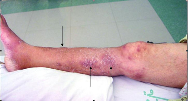
Introduction: Cutaneous mucormycosis is a rare entity related to kidney transplantation. It usually presents with ecthyma-like lesions and black necrotic cellulitis. We report an unusual case of primary cutaneous mucormycosis presenting as erythema-nodosum-like lesions in a woman who had received a renal transplant.
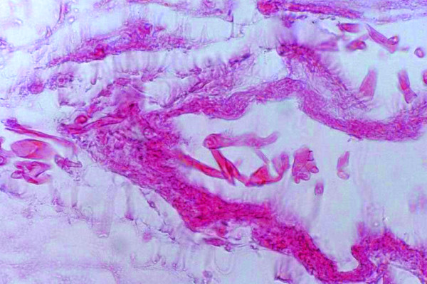
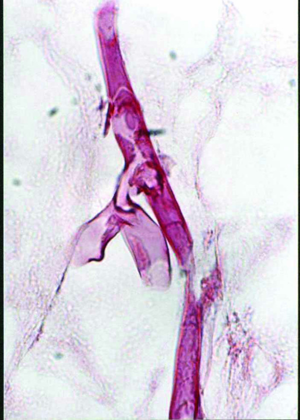
Case presentation: A 49-year-old woman with diabetes received a living-unrelated kidney transplant. Her clinical course was uneventful for the first six months after transplantation. She then developed multiple, painful, erythema-nodosum-like lesions on her right leg and thigh following an episode of minor trauma. Mucormycosis was diagnosed by skin biopsy. Microscopic examination also showed panniculitis. The patient was treated successfully with amphotericin B and surgical resection. To our knowledge, this is the first description of primary cutaneous mucormycosis with erythema-nodosum-like lesions and panniculitis after renal transplantation.
Conclusion: Cutaneous mucormycosis should be considered in the differential diagnosis when a kidney transplant recipient develops erythema-nodosum-like lesions with panniculitis.
Figures
Similar articles
-
Histoplasmosis and subcutaneous nodules in a kidney transplant recipient: erythema nodosum versus fungal panniculitis.Transpl Infect Dis. 2013 Apr;15(2):E58-63. doi: 10.1111/tid.12052. Epub 2013 Jan 20. Transpl Infect Dis. 2013. PMID: 23331504 Free PMC article.
-
Erythema nodosum migrans successfully treated with indomethacin: A rare entity.Adv Biomed Res. 2014 Dec 31;3:264. doi: 10.4103/2277-9175.148243. eCollection 2014. Adv Biomed Res. 2014. PMID: 25625103 Free PMC article.
-
Erythema nodosum in kidney transplant recipient: a rare complication of pneumonia treatment.Transpl Infect Dis. 2012 Feb;14(1):72-4. doi: 10.1111/j.1399-3062.2011.00635.x. Epub 2011 Apr 5. Transpl Infect Dis. 2012. PMID: 21466642
-
Erythema nodosum in renal transplant recipients: multiple cases and review of literature.Transpl Infect Dis. 2010 Apr;12(2):164-8. doi: 10.1111/j.1399-3062.2009.00474.x. Epub 2009 Dec 9. Transpl Infect Dis. 2010. PMID: 20002354 Review.
-
Erythema nodosum-like lesions during BRAF inhibitor therapy: Report on 16 new cases and review of the literature.J Eur Acad Dermatol Venereol. 2015 Sep;29(9):1797-806. doi: 10.1111/jdv.13039. Epub 2015 Mar 6. J Eur Acad Dermatol Venereol. 2015. PMID: 25752368 Review.
Cited by
-
Cutaneous and renal aspergillosis resulting from orthotopic liver transplantation.BMJ Case Rep. 2023 Nov 22;16(11):e256974. doi: 10.1136/bcr-2023-256974. BMJ Case Rep. 2023. PMID: 37993141 No abstract available.
-
Skin and soft tissue infections in the transplant population.Curr Infect Dis Rep. 2008 Sep;10(5):387-93. doi: 10.1007/s11908-008-0063-2. Curr Infect Dis Rep. 2008. PMID: 18687203
-
Successful treatment of a necrotizing fasciitis patient caused by Mucor indicus with amphotericin B and skin grafting.Mycopathologia. 2014 Apr;177(3-4):187-92. doi: 10.1007/s11046-014-9733-9. Epub 2014 Feb 26. Mycopathologia. 2014. PMID: 24570041
-
Global Cutaneous Mucormycosis: A Systematic Review.J Fungi (Basel). 2022 Feb 16;8(2):194. doi: 10.3390/jof8020194. J Fungi (Basel). 2022. PMID: 35205948 Free PMC article. Review.
-
Gangrenous cutaneous mucormycosis caused by Rhizopus oryzae: a case report and review of primary cutaneous mucormycosis in China over Past 20 years.Mycopathologia. 2013 Aug;176(1-2):123-8. doi: 10.1007/s11046-013-9654-z. Epub 2013 Apr 25. Mycopathologia. 2013. PMID: 23615822 Review.
References
-
- Morduchowicz G, Shmueli D, Shapira Z, Cohen SL, Yussim A, Block CS, Rosenfeld JB, Pitlik SD. Rhinocerebral mucormycosis in renal transplant recipients: report of three cases and review of the literature. Rev Infect Dis. 1986;8:441–6. - PubMed
-
- Tinmouth J, Baker J, Gardiner G. Gastrointestinal mucormycosis in a renal transplant patient. Can J Gastroenterol. 2001;15:269–71. - PubMed
-
- Ahmad M. Graft mucormycosis in a renal allograft recipient. J Nephrol. 2005;18:783–6. - PubMed
LinkOut - more resources
Full Text Sources
