. 2008 Apr 18;133(2):280-91.
doi: 10.1016/j.cell.2008.02.032.

Meiosis I is established through division-specific translational control of a cyclin

Affiliations

Meiosis I is established through division-specific translational control of a cyclin

Thomas M Carlile et al. Cell. .

Abstract

In budding yeast, key meiotic events such as DNA replication, recombination, and the meiotic divisions are controlled by Clb cyclin-dependent kinases (Clb-CDKs). Using a novel synchronization procedure, we have characterized the activity of these Clb-CDKs and observed a surprising diversity in their regulation during the meiotic divisions. Clb1-CDK activity is restricted to meiosis I, and Clb3-CDK activity to meiosis II, through 5'UTR-mediated translational control of its transcript. The analysis of cells inappropriately producing Clb3-CDKs during meiosis I furthermore defines Clb3 as an inhibitor of the meiosis I chromosome segregation program. Our results demonstrate an essential role for Clb-CDK regulation in establishing the meiotic chromosome segregation pattern.


Figures
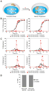
Figure 1
Figure 1. A meiotic block-release synchronization system
(A) A method for synchronizing meiotic cells using an inducible allele of NDT80. See text for details. (B and C) GAL4.ER (A14200; circles) and GAL4.ER GAL-NDT80 strains (A14201; squares) were induced to sporulate at 30°C by transfer into SPO medium. After 5 hours either ethanol (open symbols) or 1μM β-estradiol was added (closed symbols). The percentages of bi- and tri- or tetranucleate cells ([B], left graph), of tri- or tetranucleate cells ([B], right graph), and of cells with metaphase I ([C], upper left graph), anaphase I ([C], upper right graph), metaphase II ([C], lower left graph) or anaphase II spindles ([C], lower right graph) were determined at the times indicated after transfer into SPO medium. (D) GAL4.ER (A14200) and GAL4.ER GAL-NDT80 strains (A14201) were grown as described in Figure 1B. Samples were taken after 28 hours (n=40 tetrads).
Figure 2
Figure 2. Cyclin expression and activity during the meiotic divisions
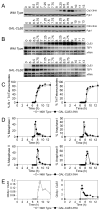
GAL4.ER GAL-NDT80 strains carrying tagged versions of cyclins CLB5-3HA (A15804), CLB4-3HA (A15090), CLB1-9Myc (A15591) and CLB3-3HA (A15802) were induced to sporulate at 30°C, and were released from the GAL-NDT80 block at 6 hours. (A, E, I and M) The amount of protein and associated Histone H1 kinase activity for the indicated cyclin ([A] Clb5-3HA, [E] Clb4-3HA, [I] Clb1-9Myc and [M] Clb3-3HA) is shown. Pgk1 or Cdc28 were used as loading controls. MetaI, AnaI, MetaII and AnaII signify the peaks of cells with metaphase I, anaphase I, metaphase II and anaphase II spindles, respectively (all time points are shown in Supplemental Figure 2A – D). (B, F, J, N) Transcript levels for the indicated cyclins ([B] CLB5-3HA, [F] CLB4-3HA, [J] CLB1-9Myc and [N] CLB4-3HA). TEF1 and rRNA are shown as loading controls. (C, G, K, O) Quantifications of cyclin transcript levels normalized to the rRNA (open symbols, left axis) and cyclin protein levels normalized to the loading control (closed symbols, right axis) for the indicated cyclins ([C] Clb5-3HA, [G] Clb4-3HA, [K] Clb1-9Myc and [O] Clb3-3HA). (D, H, L, P) Quantifications of cyclin associated Histone H1 kinase activity (open symbols, left axis) and cyclin protein levels normalized to the loading control (closed symbols, right axis) for the indicated cyclins ([D] Clb5-3HA, [H] Clb4-3HA, [L] Clb1-9Myc and [P] Clb3-3HA).
Figure 3
Figure 3. Clb3 does not accumulate during meiosis I in proteasome inhibited cells or in APC mutant cells
(A–C) Duplicate cultures of GAL4.ER GAL-NDT80 pdr5 CLB3-3HA (A16011) were sporulated at 30°C, and were released from the GAL-NDT80 block at 6 hours. Either DMSO (open symbols) or 20 μM MG-132 (closed symbols) was added at 6.5, 8.5 and 10.5 hours after transfer into SPO medium. Cells lacked the PDR5 gene to prevent export of MG-132. (D–F) Wild-type (A17946, open circles), PCLB2-CDC20 (A17855, closed circles) and PCLB2-3HA-CDC27 (A17856, open squares) cells carrying GAL4.ER, GAL-NDT80, PDS1-18Myc and CLB3-3HA alleles were sporulated at 30°C, and were released from the GAL-NDT80 block at 6 hours. (G–I) CDC23 (A17946, open symbols) or cdc23-1 (A17947, closed symbols) cells carrying GAL4.ER, GAL-NDT80, PDS1-18Myc and CLB3-3HA alleles were induced to sporulate at room temperature, were released from the GAL-NDT80 block at 7 hours, and were shifted to 34°C at 8 hours to inactivate the cdc23-1 allele. (A) Western blots for Clb3-3HA. (D, G) Western blots for Clb3-3HA and Pds1-18Myc. (B, C, E, F, H, I) The percentages of bi- and tri- or tetranucleate cells ([B, E, H], upper graphs), of tri- or tetranucleate cells ([B, E, H], lower graphs), and of cells with metaphase I ([C, F, I], upper left graphs), anaphase I ([C, F, I], upper right graphs), metaphase II ([C, F, I], lower left graphs) or anaphase II spindles ([C, F, I], lower right graphs) were determined at the times indicated after transfer into SPO medium.
Figure 4
Figure 4. The CLB3 promoter and 5’UTR are required to prevent Clb3 accumulation during meiosis I
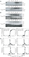
CLB3-3HA (A15055, open symbols) and GAL-CLB3-3HA (A18095, closed symbols) cells carrying GAL4.ER and GAL-NDT80 were sporulated at 30°C, and were released from the GAL-NDT80 block at 6 hours by the addition of 1 μm β-estradiol. (A) Western blots for Clb3-3HA protein. (B) Northern blots for CLB3. (C and D) The percentages of bi- and tri- or tetranucleate cells ([C], left graph), of tri- or tetranucleate cells ([C], right graph), of cells with metaphase I ([D], upper left graph), anaphase I ([D], upper right graph), metaphase II ([D], lower left graph) or anaphase II spindles ([D], lower right graph) were determined at the times indicated. (E) Quantifications of the ratio of Clb3-3HA protein to CLB3 mRNA ([Clb3p/Pgk1]/[CLB3/TEF1]). Quantifications of Clb3 protein/CLB3 RNA for wild-type are shown in the left graph. The same data was then compared with the amount of Clb3protein/CLB3 RNA generated from the GAL1-10 promoter (right graph).
Figure 5
Figure 5. The CLB3 promoter and 5’UTR are sufficient to prevent protein accumulation during meiosis I
PCLB3-CLB2 CLB3-3HA (A18574, open symbols) and PCLB4-CLB2 CLB4-3HA (A18578, closed symbols) cells carrying GAL4.ER and GAL-NDT80 alleles were cultured as described in Figure 4. (A) Western blots for Clb2, Clb3-3HA and Clb4-3HA. vATPase is shown as a loading control. (B) Northern blots for CLB2, CLB3 and CLB4. (C and D) The percentages of bi- and tri- or tetranucleate cells ([C], left graph), of tri- or tetranucleate cells ([C], right graph) and of cells with metaphase I ([D], upper left graph), anaphase I ([D], upper right graph), metaphase II ([D], lower left graph) or anaphase II spindles ([D], lower right graph) were determined at the times indicated times.
Figure 6
Figure 6. The CLB3 5’UTR is sufficient to prevent protein accumulation during meiosis I
Wild-type (A15802, open circles), GAL-CLB2 (A19060, closed circles) and GAL-5’UTRCLB3-CLB2 (A19026, open squares) cells carrying GAL4.ER, GAL-NDT80 and CLB3-3HA alleles were cultured as described in Figure 4. (A) Western blots for Clb2 and Clb3-3HA. (B) Northern blots for CLB2 and CLB3. (C and D) The percentages of bi- and tri- or tetranucleate cells ([C], upper graph), of trior tetranucleate cells ([C], lower graph), and of cells with metaphase I ([D], upper left graph), anaphase I ([D], upper right graph), metaphase II ([D], lower left graph) or anaphase II spindles ([D], lower right graph) were determined at the times indicated.
Figure 7
Figure 7. Production of Clb3 or Clb2 during meiosis I causes premature sister chromatid separation
The strains listed below were sporulated at 30°C. 1μM β-estradiol was added at 3 hrs after inoculation into SPO medium to induce CLB3 expression. (A) CLB3-3HA (A18655) and GAL-CLB3-3HA (A18656) strains carrying GAL4.ER and homozygous LEU2 dots were used. The percentage of binucleates with either one or two dots was determined at 7 hrs after inoculation into SPO ([A] left panel, n=200). The percentage of tetranucleates with one, two, three or four dots was determined at 12 hrs after inoculation into SPO ([A] right panel, n=200). (B) CLB3-3HA (A18686) and GAL-CLB3 (A18687) strains carrying GAL4.ER and heterozygous LEU2 dots, and CLB3 (A19396) and GAL-CLB3 (A19400) strains carrying GAL4.ER and heterozygous CENV dots were used. The percentage of binucleates with either one or two dots was determined at 8.5 hrs after transfer into SPO (n=200). (C) Wild Type (A19397), GAL-CLB4 (A19399) and GAL-CLB2 (A19687) strains carrying GAL4.ER and heterozygous CENV dots were used. Binucleate cells were examined 7 hrs after transfer into SPO (n=200). (D–F) CLB3 (A19402, open circles) and GAL-CLB3 (A19406, closed circles) strains carrying cdc20-mn, GAL4.ER and heterozygous CENV dots, and CLB3 (A19408, open squares) and GAL-CLB3 (A19410, closed squares) strains carrying cdc20-mn, GAL4.ER and heterozygous URA3 dots were used. The percentage of cells with separated GFP dots [D], spindles past metaphase I [E], and two DAPI masses [F] were determined at the times indicated.

References

    1. Benjamin KR, Zhang C, Shokat KM, Herskowitz I. Control of landmark events in meiosis by the CDK Cdc28 and the meiosis-specific kinase Ime2. Genes Dev. 2003;17:1524–1539. - PMC - PubMed
    1. Bloom J, Cross FR. Multiple levels of cyclin specificity in cell-cycle control. Nat Rev Mol Cell Biol. 2007;8:149–160. - PubMed
    1. Buonomo SB, Rabitsch KP, Fuchs J, Gruber S, Sullivan M, Uhlmann F, Petronczki M, Tóth A, Nasmyth K. Division of the nucleolus and its release of CDC14 during anaphase of meiosis I depends on separase, SPO12, and SLK19. Dev Cell. 2003;4:727–739. - PubMed
    1. Chu S, DeRisi J, Eisen M, Mulholland J, Botstein D, Brown PO, Herskowitz I. The transcriptional program of sporulation in budding yeast. Science. 1998;282:699–705. - PubMed
    1. Cohen-Fix O, Peters JM, Kirschner MW, Koshland D. Anaphase initiation in Saccharomyces cerevisiae is controlled by the APC-dependent degradation of the anaphase inhibitor Pds1p. Genes Dev. 1996;10:3081–3093. - PubMed

Publication types