

Mandible matrix necrosis in beagle dogs after 3 years of daily oral bisphosphonate treatment
- PMID: 18423290
- PMCID: PMC2464292
- DOI: 10.1016/j.joms.2008.01.038
Mandible matrix necrosis in beagle dogs after 3 years of daily oral bisphosphonate treatment
Abstract
Purpose: An increasing number of reports have implicated bisphosphonates as contributing to osteonecrosis of the jaw. The goal of this study was to evaluate mandible necrosis in beagle dogs treated for 3 years with oral alendronate (ALN).
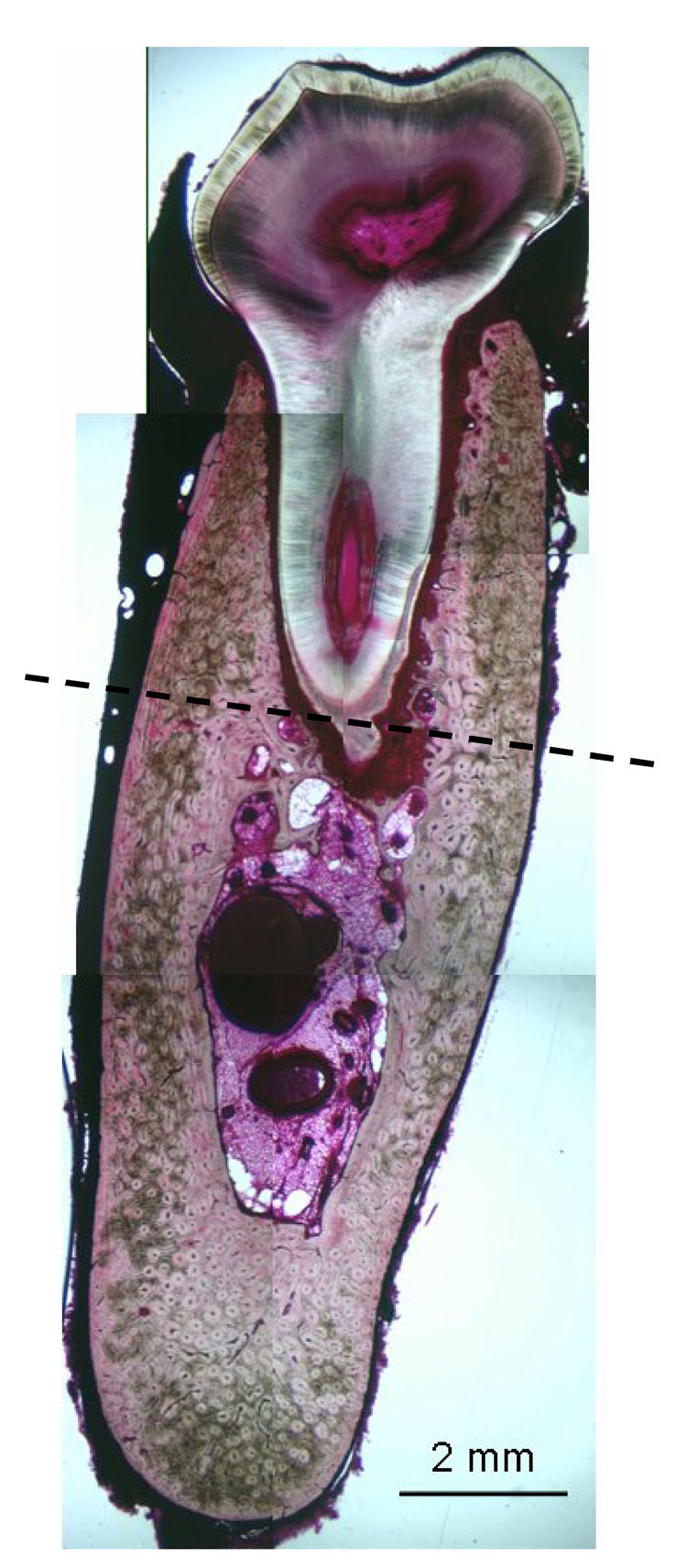
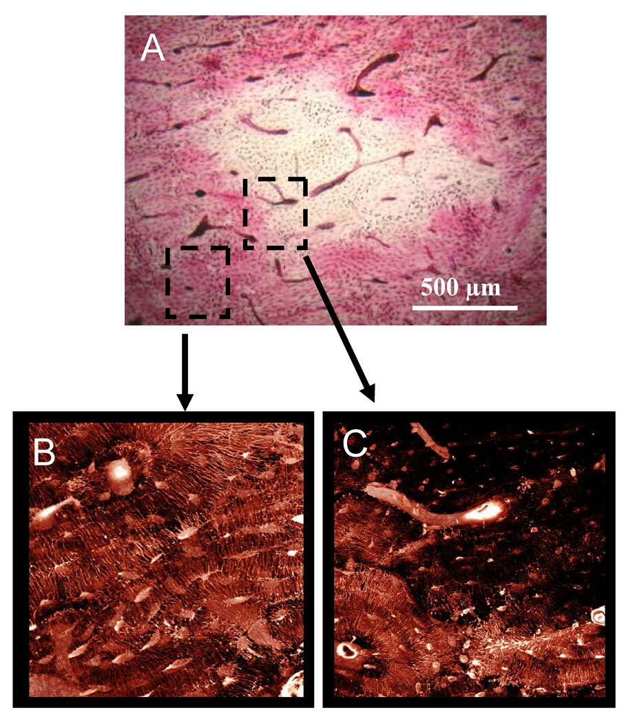
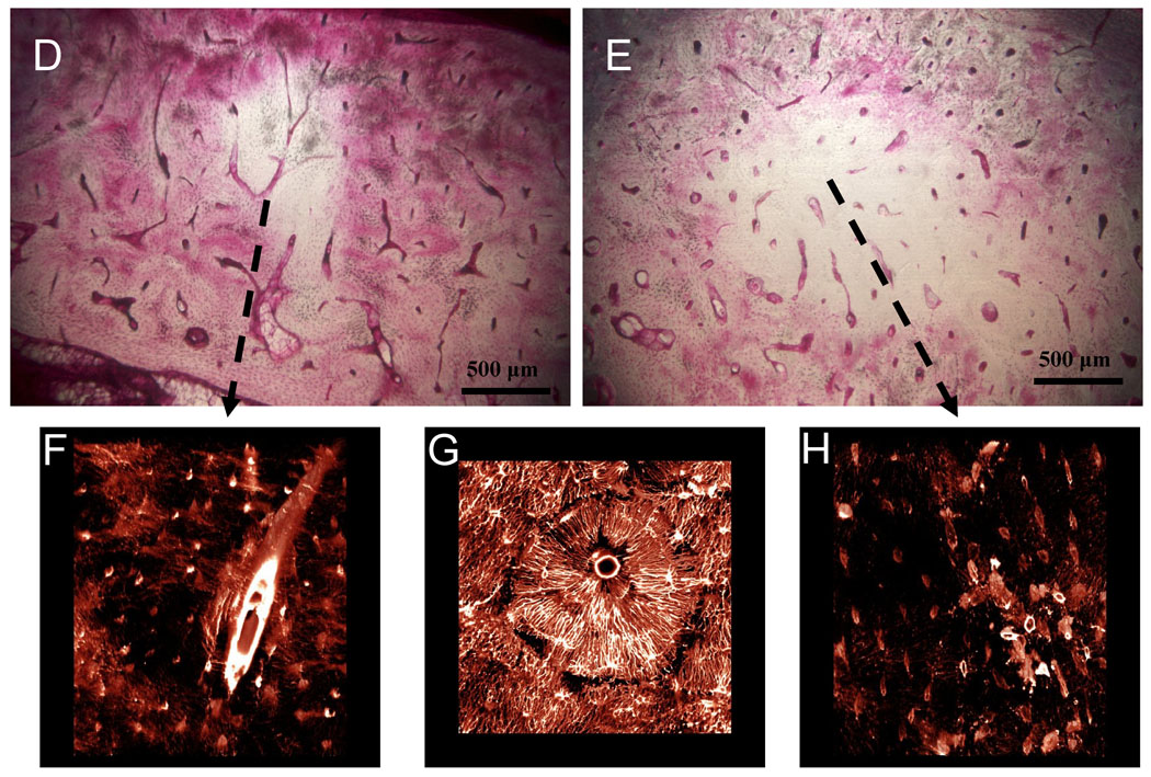
Materials and methods: Skeletally mature female beagles were treated daily for 3 years with oral doses of vehicle (VEH) or ALN (0.20 or 1.0 mg/kg/day). These doses approximate, on a mg/kg basis, those used for postmenopausal osteoporosis and Paget's disease, respectively. At necropsy, the second molar region of the mandible was excised, stained en bloc with basic fuchsin, and assessed for matrix necrosis and intracortical bone turnover rate using histology. Matrix necrosis was defined as a region greater than 500 microm(2) that was void of basic fuchsin stain, assessed using both bright-field and confocal microscopy.
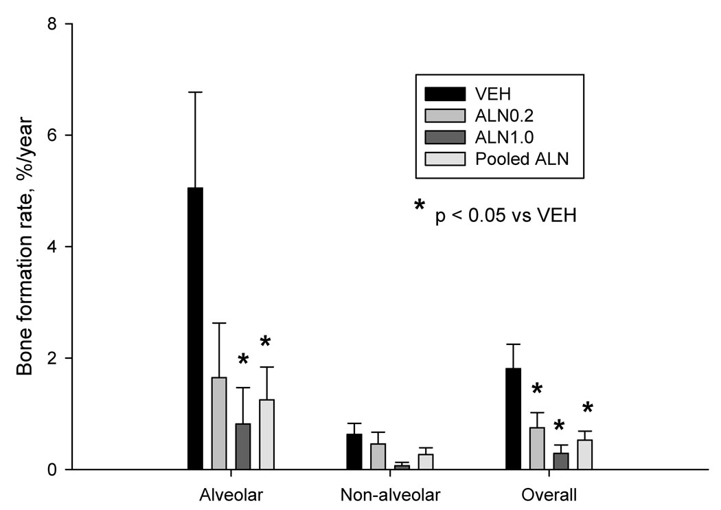
Results: No animals developed exposed bone lesions in the oral cavity during the 3-year study. Matrix necrosis was observed in 25% of ALN0.2 animals, 33% of ALN1.0 animals, and was noticeably absent from all vehicle animals (P < .05 pooled ALN doses vs VEH). These necrotic regions occurred predominately in the alveolar bone and were clearly void of patent canaliculi. Intracortical bone turnover rate of the alveolar mandible bone region was significantly lower (-75%, P < .05) in ALN-treated animals compared with VEH.
Conclusions: Three years of daily oral bisphosphonate treatment reduces bone turnover significantly and increases the incidence of matrix necrosis within the mandible of dogs.
Figures
References
-
- Enlow DH. Functions of the Haversian system. Am J Anat. 1962;110:269–305. - PubMed
-
- Enlow DH. Osteocyte necrosis in normal bone. J Dent Res. 1966;45:213. - PubMed
-
- Frost HM. In vivo osteocyte death. J Bone Joint Surg Am. 1960;42-A:138–143. - PubMed
-
- Dunstan CR, Somers NM, Evans RA. Osteocyte death and hip fracture. Calcif Tissue Int. 1993;53 Suppl 1:S113–S116. discussion S116-117. - PubMed
-
- Migliorati CA, Casiglia J, Epstein J, et al. Managing the care of patients with bisphosphonate-associated osteonecrosis: an American Academy of Oral Medicine position paper. J Am Dent Assoc. 2005;136:1658–1668. - PubMed
Publication types
MeSH terms
Substances
Grants and funding
LinkOut - more resources
Full Text Sources
Medical
Research Materials
