. 2008 Jun 1;318(1):1-16.
doi: 10.1016/j.ydbio.2008.02.022. Epub 2008 Mar 4.

Polychaetoid controls patterning by modulating adhesion in the Drosophila pupal retina

Affiliations

Polychaetoid controls patterning by modulating adhesion in the Drosophila pupal retina

Midori J Seppa et al. Dev Biol. .

Abstract

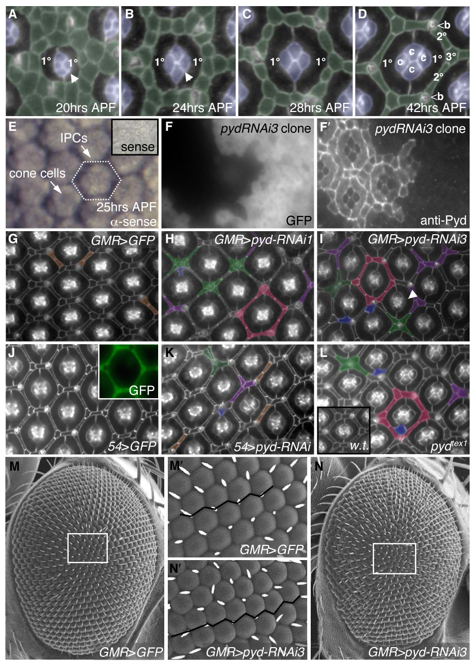
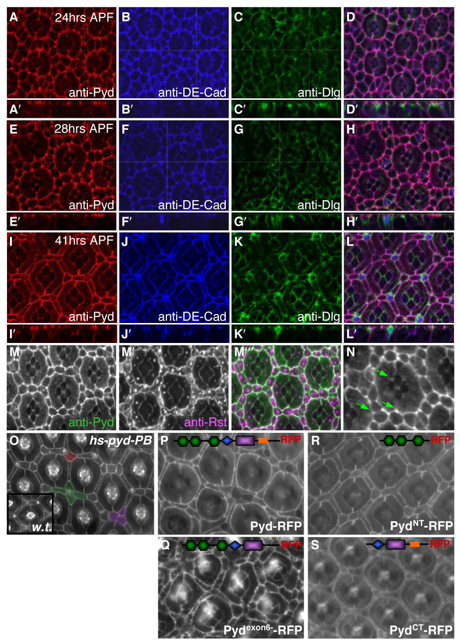
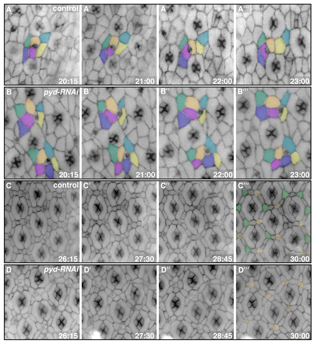
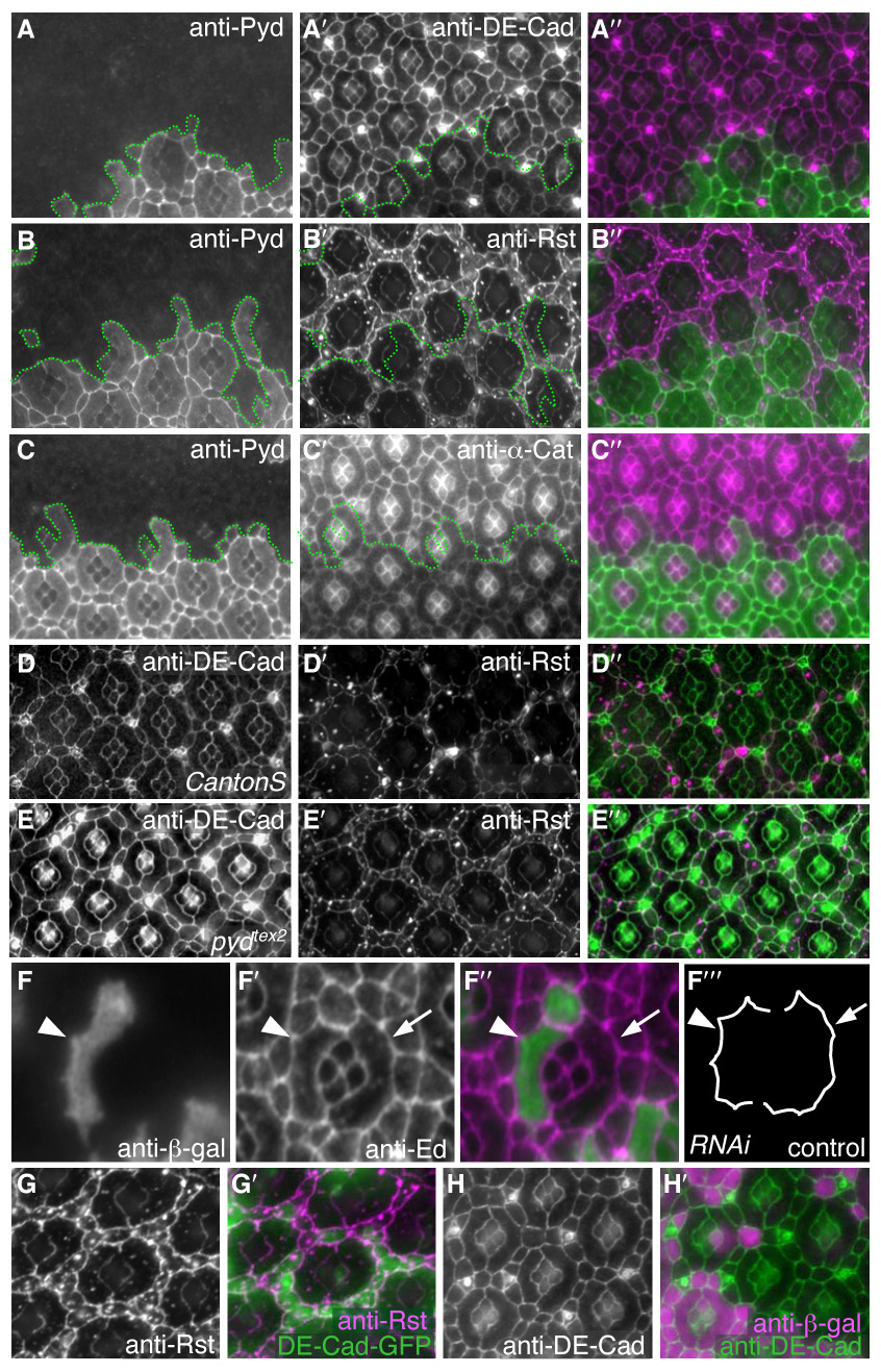
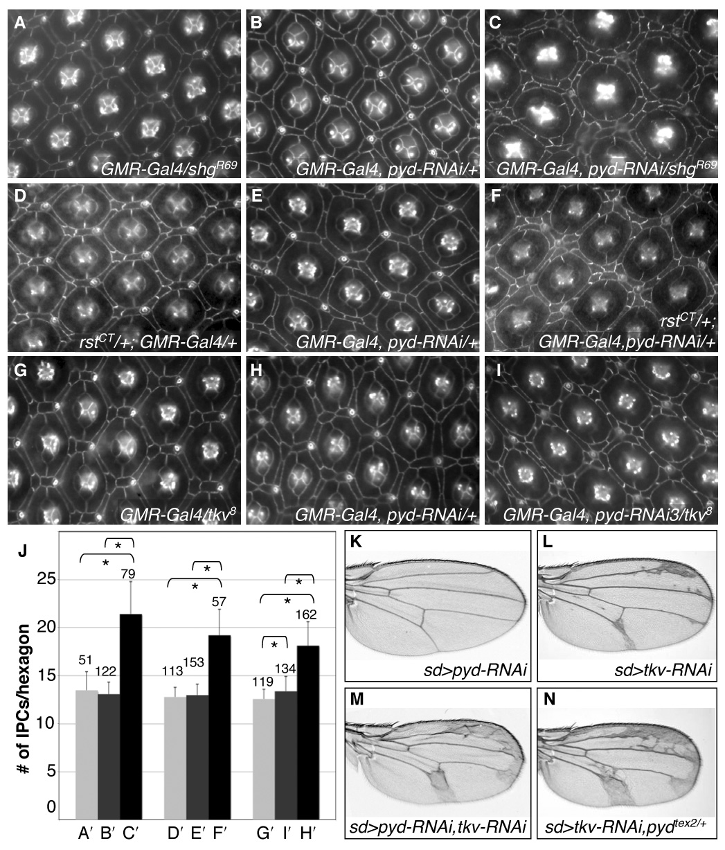
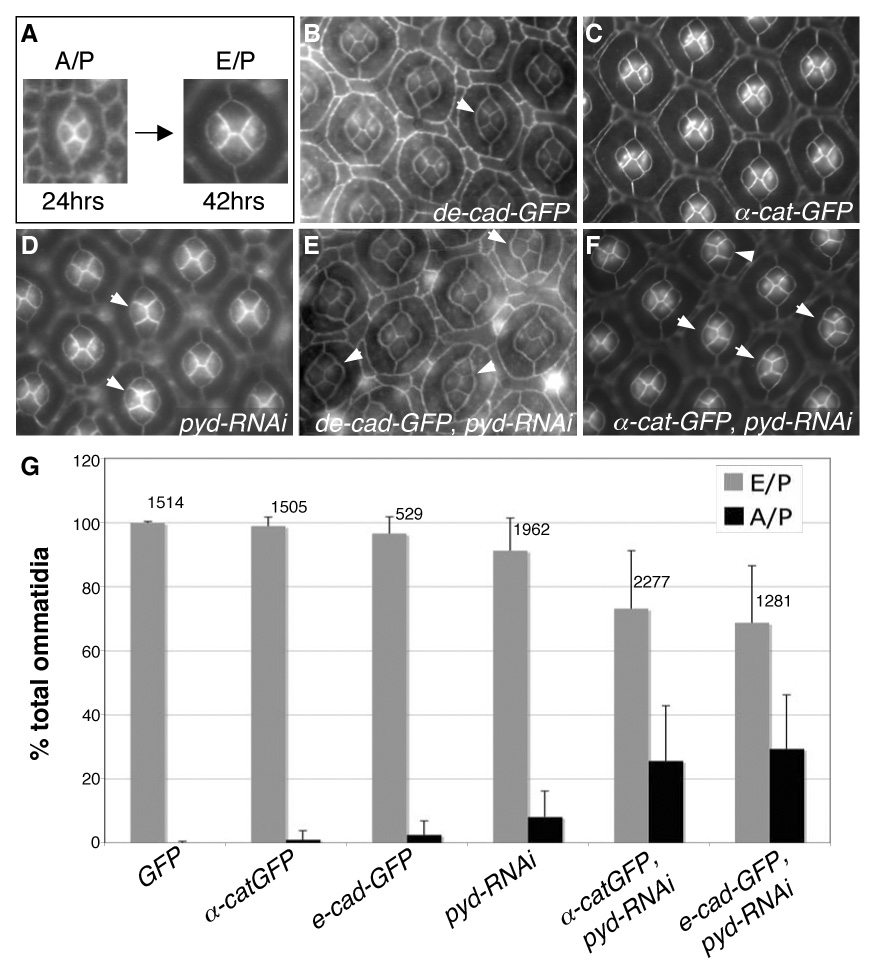
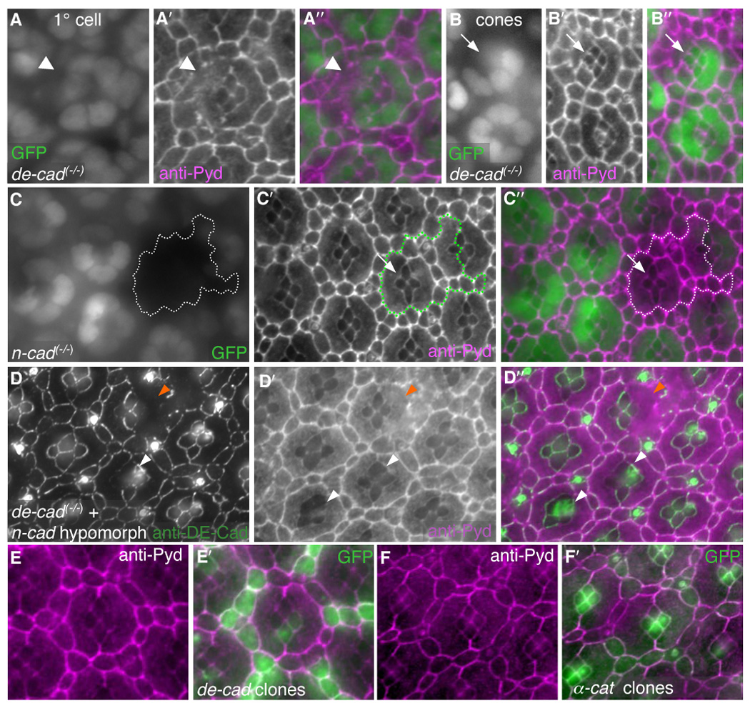
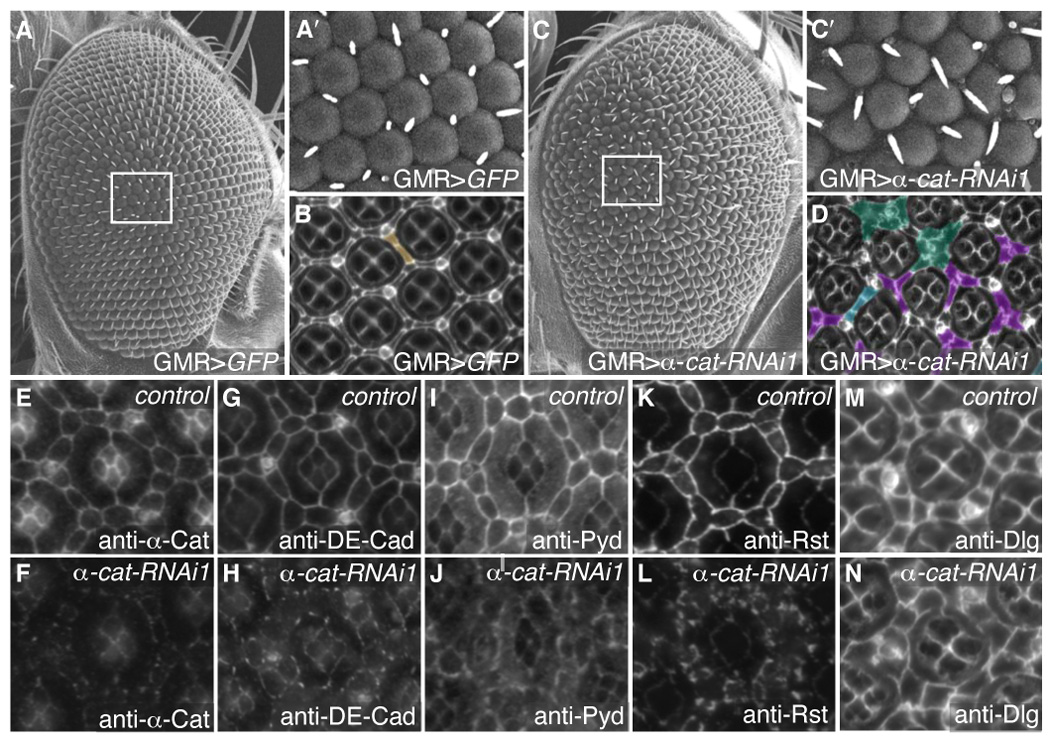
Correct cellular patterning is central to tissue morphogenesis, but the role of epithelial junctions in this process is not well-understood. The Drosophila pupal eye provides a sensitive and accessible model for testing the role of junction-associated proteins in cells that undergo dynamic and coordinated movements during development. Mutations in polychaetoid (pyd), the Drosophila homologue of Zonula Occludens-1, are characterized by two phenotypes visible in the adult fly: increased sensory bristle number and the formation of a rough eye produced by poorly arranged ommatidia. We found that Pyd was localized to the adherens junction in cells of the developing pupal retina. Reducing Pyd function in the pupal eye resulted in mis-patterning of the interommatidial cells and a failure to consistently switch cone cell contacts from an anterior-posterior to an equatorial-polar orientation. Levels of Roughest, DE-Cadherin and several other adherens junction-associated proteins were increased at the membrane when Pyd protein was reduced. Further, both over-expression and mutations in several junction-associated proteins greatly enhanced the patterning defects caused by reduction of Pyd. Our results suggest that Pyd modulates adherens junction strength and Roughest-mediated preferential cell adhesion.


Figures

Fig 1
Fig 1
pyd regulates patterning of IPCs. Apical cell profiles were detected with anti-Arm antibodies at 41–42 hours APF (after puparium formation) unless otherwise indicated. Anterior is to the right in this and all subsequent images. A–D. Normal development of the pupal eye is marked by sorting of the IPCs (pseudo-colored in green) into a hexagonal lattice. Between 18–24 hours APF, cell intercalation narrows the rows of IPCs to single file (B). The hexagon is composed of six 2°s forming the sides of the hexagon and three 3°s at the vertices, alternating with bristles (b) (D). E–E′. In situ hybridization using full-length pyd-RB anti-sense probe in 25 hr APF pupal retinas: pyd was expressed at highest levels in IPCs and at lower levels in cone cells and 1°s. Inset shows pyd-RB sense probe. F–F′. Expression of pyd-RNAi3 in clonal patches in the eye marked by GFP (F) demonstrated cell-autonomous loss of Pyd antibody staining at the apical membrane (F′). G–L. Examples of patterning defects are pseudo-colored as follows: extra cells in the absence of other defects (brown), clustering of cells around bristles (green), pentagonal ommatidia (red), IPC-cone contact cells (blue) and a failure to specify a single 3° cell (purple). G. Control GMR>GFP retinas. H–I. GMR>pyd-RNAi1 (H) or GMR>pyd-RNAi3 (I) resulted in far stronger IPC patterning defects. Note also the failure to switch cone-cell contacts (I; arrowhead). J. 54>GFP retinas have no patterning defects (J). Expression of 54-Gal4, visualized with GFP, is found primarily in IPCs (inset). K. 54>pydRNAi exhibited errors in IPC organization. L. pydtex1 mutant eyes had defects that were similar to RNAi expression; inset shows wild-type retina. M–N. SEM of control (M) and pyd-RNAi3 (N) expressing eyes. The white tracing outlines an area that is shown at higher magnification in M′ and N′, respectively. The black line emphasizes the slightly uneven ommatidial rows in pyd-RNAi expressing eyes (N′) vs. control (M′).
Fig 2
Fig 2
Pyd localized to AJs and its over-expression led to pupal eye patterning defects. A–L. Apical confocal sections of wild type pupal eye were taken at 24 hours APF (A–D), 28 hours APF (E–H) and 41 hours APF (I–L). Anti-Pyd immunofluorescence (A, E, I) co-localized extensively with anti-DE-Cad (B, F, J) at the AJ but only overlapped slightly with the septate junction marker Dlg (C, G, K). D, H, L. Overlay of anti-Pyd, anti-DE-Cad and anti-Dlg. Inset panels A′–L′ are lateral projections. M–M″. Anti-Pyd (M) co-localized with anti-Rst (M′) at the IPC/1° interface at 28 hours APF (overlay in M″). N. Anti-Pyd was concentrated where three or more cells came together (‘tricellular junctions’; green arrows). O. 41 hours APF pupal CS (inset) or hs-pyd-RB (O) eyes with a 30 min 37°C heat-shock at 17 hours APF. Notice that some phenotypes were shared with pyd-RNAi, such as the clustering of cells around bristles (pseudo-colored green) and the failure to specify a single 3° cell (pseudo-colored purple). Occasionally, a 2° extended to cover both the 2° and 3° niches (pseudo-colored red). P–S. GMR>UAS-pyd-RFP expression in 42 hr APF eyes; the relevant domains of each construct are schematized (see Supp Fig 3A). Pyd-RFP (P) localized to the AJ and directed patterning defects similar to those observed with pyd-RNAi expression. Pydexon6--RFP localized to the AJ and had severe cone cell and IPC defects (Q). PydNT-RFP and PydCT-RFP localized to the AJ but both rarely exhibited patterning defects (R and S, respectively).
Fig 3
Fig 3
pyd-RNAi expressing cells failed to execute productive movements or sort appropriately. Membranes were labeled with α-Catenin-GFP. Times represent hours APF. A–B‴. Panels from four time points during cell intercalation for both control (A–A‴) and pyd-RNAi (B–B‴) retinas. Cells were pseudo-colored to emphasize particular cell movements. Typically, pyd-RNAi-expressing cells failed to undergo intercalation, instead remaining in double rows. C–D‴. Panels from four time points during 3° cell patterning for both control (C–C‴) and pyd-RNAi (D–D‴) retinas. Control cells moved into and out of the 3° position early; by 30 hours, however, one cell had taken over the niche in most cases (green in C‴; bristles were pseudo-colored in orange in C‴ and D‴). By contrast, the pyd-RNAi-expressing cells maintained initial contacts and, in most cases, failed to establish a single 3° (D‴).
Fig 4
Fig 4
Cells with reduced Pyd exhibited increased levels of core AJ proteins at the membrane. All images were taken at 28 hours APF. A–C″. pyd-RNAi expressing cells were marked by lack of anti-Pyd immunofluorescence (A, B, C). DE-Cad (A′), Rst (B′) and α-Cat (C′) levels were increased specifically at the apical membrane in cells expressing pyd-RNAi. Overlay in A″, B″ and C″, respectively. D–E″. Homozygous pydtex2 retinas also had increased levels of DE-Cad (E) and Rst (E′) – compare to wild type tissue (D and D′). Overlay in D″ and E″. F–F‴. Single 1° cell and IPC clones expressing pyd-RNAi were marked by β-gal expression (F). Anti-Echinoid (F′) outlines the cells (overlay in F″). Tracing of control and pyd-RNAi expressing cells (F‴) demonstrating that scalloping of the IPC/1° boundary (e.g., arrowhead) was increased when cells expressed pyd-RNAi. G–G′. DE-Cad-GFP over-expression in a patch of cells (green in G′) did not alter the localization or level of anti-Rst immunofluorescence (G and magenta in G″). H–H′. Rst over-expression in a patch of cells (marked by anti-β-gal; magenta in H′) did not alter the localization or level of anti-DE-Cad (green in H″).
Fig 5
Fig 5
pyd is functionally linked to AJ proteins involved in adhesion. A–I. Control GMR-Gal4/shgR69 (A), rstCT/+; GMR-Gal4/+(D) and GMR-Gal4/tkv8 retinas exhibited few IPC patterning errors. GMR>pyd-RNAi retinas (B, E, H) had mild IPC patterning errors. GMR>pyd-RNAi defects were strongly enhanced by removal of one functional copy of de-cad/shg (C; GMR-Gal4, pyd-RNAi/shgR69), rst (F; rstCT/+; GMR-Gal4, pyd-RNAi/+) or the Dpp pathway Type I receptor tkv (I; GMR-Gal4, pyd-RNAi/tkv8). J. Number of IPCs per hexagonal area (see Methods) for each genotype, respectively. Error bars represent standard deviation, N numbers are given above each bar. Brackets with stars represent statistically significant differences at p<0.001 by the Mann-Whitney U test. K–N. Reducing Pyd with the wing driver scalloped-Gal4 (sd>pyd-RNAi) led to no visible effect on the wing veins (K). Reducing Tkv protein levels using scalloped-Gal4 (sd>tkv-RNAi) resulted in mild expansion of the wing vein material (L). When Pyd and Tkv were jointly reduced (sd>pyd-RNAi, tkv-RNAi) the wing vein phenotype was strongly enhanced (M). Similarly the tkv-RNAi phenotype was greatly enhanced in pydtex heterozygotes (N). All wings are from females.
Fig 6
Fig 6
pyd regulates the A/P to E/P cone cell contact switch. A. Between 24–28 hours APF, cone cells switch their apical contacts from an anterior/posterior (A/P) to an equatorial/polar (E/P) dominant configuration. B–G. GMR>pyd-RNAi expression interfered with this switching, and a subset of cone cell groups maintained the immature A/P arrangement through 42 hours APF (D, arrows). Expression of either UAS-de-cad-GFP (B, arrow) or UAS-α-cat-GFP (C) during pupal development resulted in a low level failure to switch cone cell-contacts. Expression of UAS-de-cad-GFP or UAS-α-cat-GFP strongly enhanced the pyd-RNAi cone cell switching phenotype (arrowheads in E and F, respectively). Data from the relevant genotypes is quantified in G; error bars represent standard deviation for each eye field, N numbers are given above each set of bars.
Fig 7
Fig 7
DE-Cad is necessary for Pyd to localize to the AJ. All images taken at 28 hours APF. A–B′. Single-cell null de-cad/shotgun (shg1H) 1° clones (A, arrowhead) and cone cell clones (B, arrow) are marked by loss of nuclear GFP expression. In isolated 1° cell clones (A), Pyd de-localized from the membranes of the adjoining IPC and cone cells that contacted the de-cad deficient cell membrane (A′, arrowhead). Pyd was maintained at the cone/cone interface in de-cad null cone cell clones (B′, arrow). C–C″. Null n-cadherin (n-cadM19) clones, marked by lack of nuclear GFP (C), showed no change in Pyd localization (C′) at the cone/cone interface (arrow, overlay in C″). D–D″. Null de-cad (shgR69) single-cell clones (marked by loss of DE-Cad staining in D) in the background of a strong reduction in n-cadherin (n-cadM12/n-cadM19) exhibited de-localized Pyd staining at the cone/cone interface (D′, white arrowheads). Null de-cad clones in 1°s were also observed (orange arrrowheads). D″ shows overlay. E–E″. DE-Cad-over-expression in single cells (labeled with GFP, green in E′) did not alter the localization or level of anti-Pyd immunofluorescence (E and magenta in E′). F–F′. UAS-α-catenin expression in single cells (labeled with GFP, F′) did not alter the localization or level of anti-Pyd immunofluorescence (F and magenta in F′).
Fig 8
Fig 8
α-Catenin is necessary for AJ protein localization. A–A′. SEM of control GMR>GFP adult eyes demonstrating even ommatidial rows (A) with boxed area shown enlarged in A′. B. 41 hours APF pupal eye stained with anti-Discs large to mark cell membranes. Extra cell was pseudo-colored in brown. C–C′. Reduction of α-Catenin (GMR>α-cat-RNAi) disrupted the orderly array of ommatidial rows in the adult eye (C). C′ shows an enlargement of the boxed area C. D. 41 hours APF pupal eye stained with anti-Discs large and pseuso-colored to show examples of patterning errors: extra cells (blue), clustering of IPCs around bristles (green), and the failure of only one cell to occupy the 3° niche (purple). E–N. 28 hours APF pupal eyes. E–F. GMR>α-cat-RNAi retinas showed greatly reduced levels of α-Catenin (F) compared to GMR>GFP expressing eyes (E): the image in F represents >8-fold longer exposure than E. G–N. Control GMR>GFP retinas visualizing DE-Cad (G), Pyd (I), Rst (K) and Dlg (M). Expression of GMR>α-cat-RNAi in the pupal retina resulted in de-localization of DE-Cad (H), Pyd (J) and Rst (L), but no change in anti-Dlg staining (N).

References

    1. Aijaz S, Balda MS, Matter K. Tight junctions: molecular architecture and function. Int Rev Cytol. 2006;248:261–298. - PubMed
    1. Bao S, Cagan R. Preferential adhesion mediated by Hibris and Roughest regulates morphogenesis and patterning in the Drosophila eye. Dev Cell. 2005;8:925–935. - PubMed
    1. Bao S, Cagan R. Fast cloning inverted repeats for RNA interference. Rna. 2006;12:2020–2024. - PMC - PubMed
    1. Barletta GM, Kovari IA, Verma RK, Kerjaschki D, Holzman LB. Nephrin and Neph1 co-localize at the podocyte foot process intercellular junction and form cis hetero-oligomers. J Biol Chem. 2003;278:19266–19271. - PubMed
    1. Brachmann CB, Cagan RL. Patterning the fly eye: the role of apoptosis. Trends Genet. 2003;19:91–96. - PubMed

Publication types

MeSH terms

LinkOut - more resources