. 2008 Jun 20;376(1):42-52.
doi: 10.1016/j.virol.2008.03.007. Epub 2008 Apr 18.

Nuclear trafficking of the human cytomegalovirus pp71 (ppUL82) tegument protein

Affiliations

Nuclear trafficking of the human cytomegalovirus pp71 (ppUL82) tegument protein

Weiping Shen et al. Virology. .

Abstract

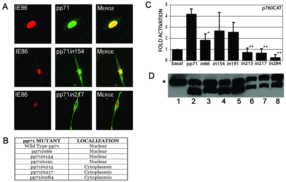
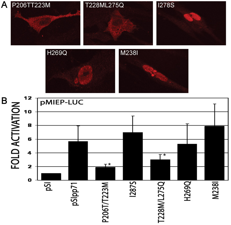
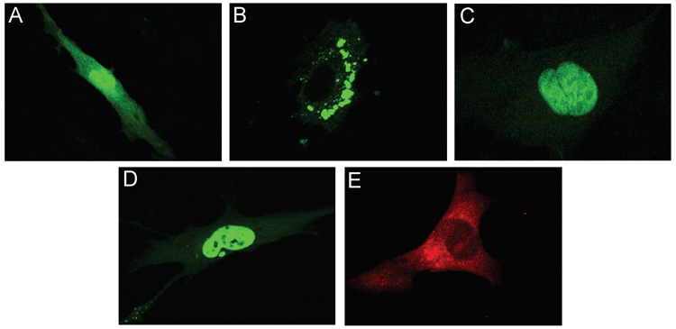
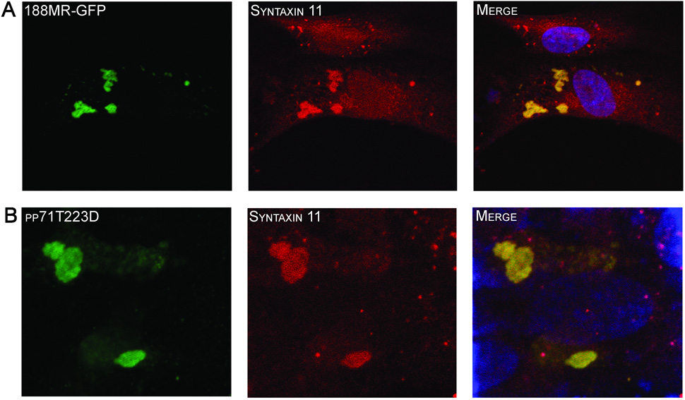
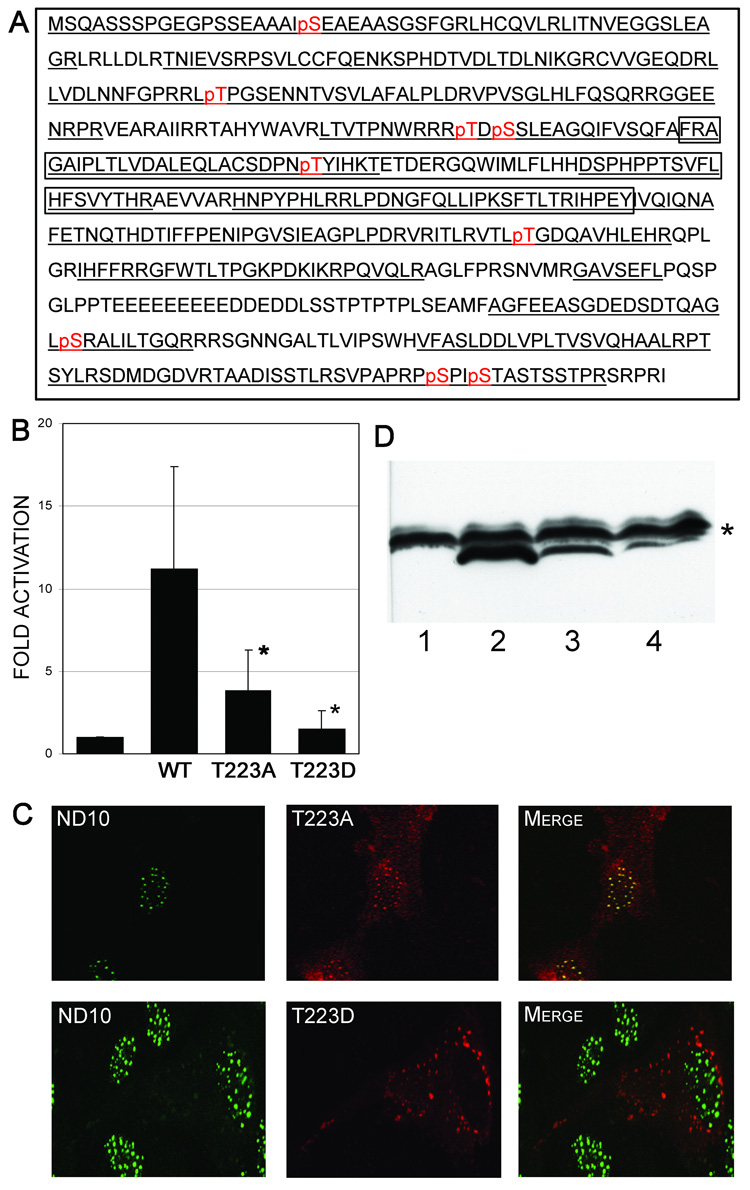
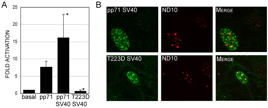
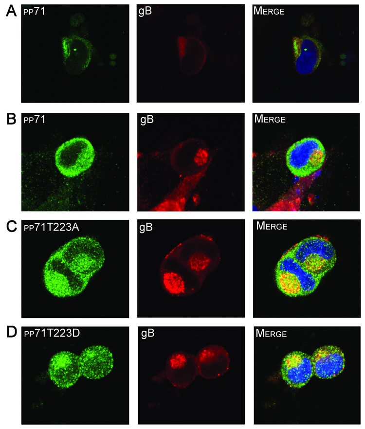
The human cytomegalovirus tegument protein pp71 localizes to the nucleus immediately upon infection, and functions to initiate viral gene expression. Analysis of a series of random insertion mutations revealed that sequences within the mid region (MR) of pp71 are important for localization to the nucleus. Fusion of MR sequences with eGFP revealed that amino acids 94 to 300 were sufficient to target proteins to the nucleus. Random substitution mutagenesis within this domain resulted in two double substitution mutants, pp71P203T/T223M and pp71T228M/L275Q, with a predominantly cytoplasmic localization. Disruption of nuclear targeting resulted in relocalization of the fusion proteins to a distinct perinuclear region. Using tandem mass spectrometry, we determined that threonine 223 can be phosphorylated. Mutation of this residue to a phosphomimetic amino acid resulted in abrogation of nuclear targeting. These results strongly suggest that the intracellular trafficking of pp71 is regulated by phosphorylation.


Figures

FIGURE 1
FIGURE 1
A & B. Subcellular localization of pp71 insertion mutants. HFF cells were co-transfected with constructs expressing the indicated pp71 insertion mutants and pMCRS86, and processed for immunofluorescence and confocal microscopy 48 hours after transfection using primary antibodies to the HA epitope tag of pp71 and IE86, with goat anti-mouse Alexa Fluor® 488 and anti-rabbit Alexa Fluor® 546 as secondary antibodies. The pp71 proteins are shown as green fluorescence, and IE86 shown as red fluorescence. The pp71in154 and pp71in217 proteins are shown as representatives of the insertion mutant phenotypes as indicated in B. C. Transcriptional activity of pp71 insertion mutants. HFF cells were co-transfected with p760-CAT and pCMV71HAX or the indicated insertion mutants. Cells were harvested 48 hours post-transfection and assessed for CAT activity. Data is the average and standard deviation of a minimum of three experiments, normalized to promoter activity in the absence of pp71. Symbols: * significantly different from wild type, p<0.01; ** significantly different from wild type, p<0.001. D. Western blot analysis of the pp71 insertion mutants. HeLa cells were transfected with the following plasmids: pSI (lane 1), pSI71HAX (lane 2), pSI71HAXin66 (lane 3), pSI71HAXin154 (lane 4), pSI71HAXin191 (lane 5), pSI71HAXin215 (lane 6), pSI71HAXin217 (lane 7) or pSI71HAXin284 (lane 8). Cells were harvested at 48 hours post-transfection and the extracts subjected to western blot analysis using an antibody to the HA-epitope tag. Symbol: * non-specific band used as a loading control.
FIGURE 2
FIGURE 2
A. Subcellular localization of pp71 mutants generated by random mutagenesis. HFF cells were transfected with constructs expressing the indicated pp71 mutants, and processed for immunofluorescence and confocal microscopy 48 hours after transfection using antibodies to the HA epitope tag. B. Transcriptional activity of pp71 mutants. HFF cells were transfected with pMIEP-LUC and pSI71HAX or the indicated mutants. Cells were harvested 48 hours post-transfection and assessed for luciferase activity. Data is the average and standard deviation of a minimum of three experiments, normalized to promoter activity in the absence of pp71. Symbol: * significantly different from wild type, p<0.05.
FIGURE 3
FIGURE 3. Subcellular localization of MR-eGFP fusion proteins
HFF cells were transfected with peGFP-N2 (A), p188MR-eGFP (B), p94MR-eGFP (C), or p62MR-eGFP (D) and processed for confocal microscopy 48 hours after transfection. HFF cells infected with HCMV were processed for immunofluoresence at 72 hours post-infection and subjected to immunofluorescence using a monoclonal antibody to pp71 (E).
FIGURE 4
FIGURE 4. Characterization of pp71 cytoplasmic localization
HFF cells were transfected constructs expressing p188MR-eGFP (A) or pp71T223D (B) and processed for confocal microscopy 48 hours after transfection. Cells transfected with the construct expressing pp71T223D were stained using and antibody to the HA-epitope tag (green) and both were costained with an antibody to syntaxin 11 (red). Nuclei were stained with TOTO3 dye (blue).
FIGURE 5
FIGURE 5
A. Phosphopeptide map of pp71. A compilation of the results obtained with MS/MS analysis of phosphorylation sites of the pp71 tegument protein. The total analyzed sequence is underlined, and the phosphorylated residues are indicated in red. The region corresponding to the Mid-Region (MR, aa 200–300) is boxed. B. Transcriptional activity of pp71 phosphorylation site mutants. HFF cells were transfected with pMIEP-LUC and pSI71HAX or the indicated mutants. Cells were harvested 48 hours post-transfection and assessed for luciferase activity. Data is the average and standard deviation of a minimum of three experiments and is expressed relative to the activity of the wild type (WT) pp71 protein. Symbol: * significantly different from wild type, p<0.05. C. Subcellular localization of pp71 phosphorylation site mutants. HFF cells were transfected with constructs expressing the indicated pp71 mutants, and processed for immunofluorescence and confocal microscopy 48 hours after transfection using antibodies to the HA epitope tag and nuclear ND10 domains. The pp71 proteins are shown as red fluorescence, and ND10 domains shown as green fluorescence. D. Western Blot analysis of pp71 phosphorylation site mutants. HeLa cells were transfected with constructs expressing eGFP (lane 1), pp71 (lane 2), pp71T223A (lane 3), or pp71T223D (lane 4). Cells were harvested at 48 hours post-transfection and the extracts subjected to western blot analysis using an antibody to the HA-epitope tag. Symbol: * non-specific band used as a loading control.
FIGURE 6
FIGURE 6
A. Transcriptional activity of pp71 mutants containing the SV40 NLS. HFF cells were transfected with pMIEP-LUC and pSI71HAX or the indicated mutants. Cells were harvested 48 hours post-transfection and assessed for luciferase activity. Data is the average and standard deviation of a minimum of three experiments and is expressed relative to the activity of the wild type (WT) pp71 protein. Symbol: * significantly different from wild type, p<0.01. B. Subcellular localization of pp71 mutants containing the SV40 NLS. HFF cells were transfected with constructs expressing the indicated pp71 mutants, and processed for immunofluorescence and confocal microscopy 48 hours after transfection using antibodies to the HA epitope tag and nuclear ND10 domains. The pp71 proteins are shown as green fluorescence, and ND10 domains shown as red fluorescence.
FIGURE 7
FIGURE 7. Subcellular localization of pp71 at the late stages of virus infection
HFF cells were infected with HCMV strain AD169 (A) and fixed at 96 hours post-infection. The cells were then subjected to immunofluorescence using a monoclonal antibody to pp71 to or a polyclonal antibody to gB. Alternatively, HFF cells were transfected with constructs expressing pp71 (B), pp71T223A (C), or pp71T223D (D) and subsequently infected with HCMV strain AD169. Cells were fixed at 96 hours post-infection and subjected to immunofluorescence using an antibody to the HA epitope tag of pp71 (green) and the gB protein (red). Nuclei were stained with TOTO3 dye (blue).

References

    1. Abate DA, Watanabe S, Mocarski ES. Major human cytomegalovirus structural protein pp65 (ppUL83) prevents interferon response factor 3 activation in the interferon response. J Virol. 2004;78(20):10995–11006. - PMC - PubMed
    1. Anders DG, Kerry JA, Pari GS. Betaherpesviruses: DNA synthesis and late viral gene expression. In: Arvin A, Campadielli-Fume G, Mocarski ES, Moore P, Roizman B, Whitley R, Yamanishi K, editors. "Human Herpesviruses: Biology, Therapy and Immunoprophylaxis". Cambridge: Cambridge University Press; 2007. pp. 295–310.
    1. AuCoin DP, Smith GB, Meiering CD, Mocarski ES. Betaherpesvirus-conserved cytomegalovirus tegument protein ppUL32 (pp150) controls cytoplasmic events during virion maturation. J Virol. 2006;80(16):8199–8210. - PMC - PubMed
    1. Baldick CJ, Jr, Marchini A, Patterson CE, Shenk T. Human cytomegalovirus tegument protein pp71 (ppUL82) enhances the infectivity of viral DNA and accelerates the infectious cycle. J Virol. 1997;71(6):4400–4408. - PMC - PubMed
    1. Baldick CJ, Jr, Shenk T. Proteins associated with purified human cytomegalovirus particles. J Virol. 1996;70(9):6097–6105. - PMC - PubMed

Publication types

MeSH terms

Substances