. 2008 May;82(5):1178-84.
doi: 10.1016/j.ajhg.2008.03.007.

Mutation in the human homeobox gene NKX5-3 causes an oculo-auricular syndrome

Affiliations

Mutation in the human homeobox gene NKX5-3 causes an oculo-auricular syndrome

Daniel F Schorderet et al. Am J Hum Genet. 2008 May.

Abstract

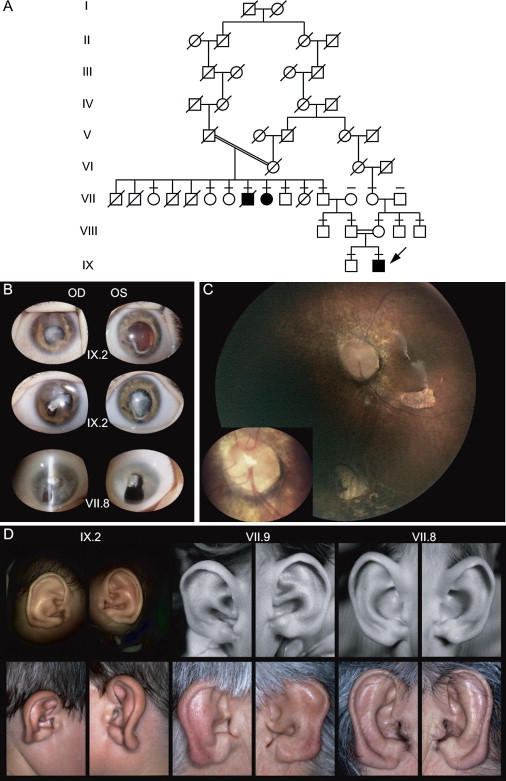
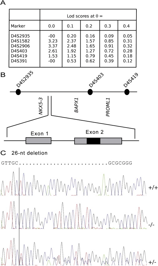
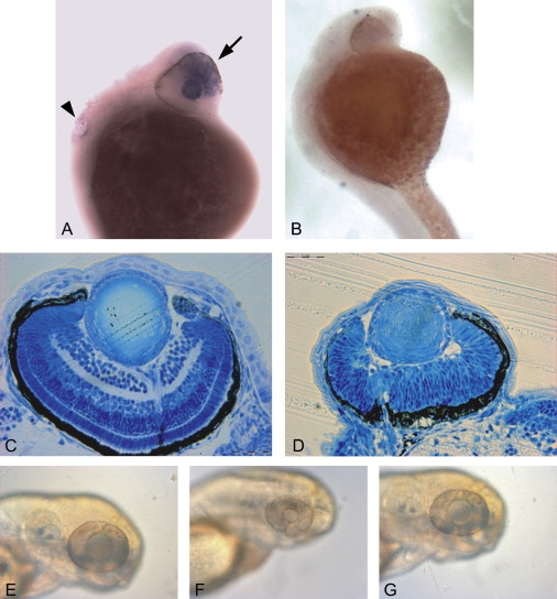
Several dysmorphic syndromes affect the development of both the eye and the ear, but only a few are restricted to the eye and the external ear. We describe a developmental defect affecting the eye and the external ear in three members of a consanguineous family. This syndrome is characterized by ophthalmic anomalies (microcornea, microphthalmia, anterior-segment dysgenesis, cataract, coloboma of various parts of the eye, abnormalities of the retinal pigment epithelium, and rod-cone dystrophy) and a particular cleft ear lobule. Linkage analysis and mutation screening revealed in the first exon of the NKX5-3 gene a homozygous 26 nucleotide deletion, generating a truncating protein that lacked the complete homeodomain. Morpholino knockdown expression of the zebrafish nkx5-3 induced microphthalmia and disorganization of the developing retina, thus confirming that this gene represents an additional member implicated in axial patterning of the retina.


Figures

Figure 1
Figure 1
Family with Oculo-Auricular Syndrome (A) Pedigree of consanguineous family with autosomal-recessive oculo-auricular syndrome. Affected individuals are indicated by black symbols, and the proband is indicated by an arrow. Individuals with line above symbol have been tested for mutation. (B) Eyes of two affected patients. Top: Eyes of the proband (IX.2) showing an early cataract, coloboma, and microphthalmia of OD (15.5 mm); OS was borderline (16.5 mm). At age 7 yr, best corrected visual acuity was 0.05 in OD and 0.16 in OS; corneal thickness was 770 μm OD and 710 μm OS because of a diffuse cornea guttata. Middle: Eyes of the proband a few months later with advanced cataract. Bottom: Eyes of VII.8 presenting cataract and coloboma. (C) Fundus photography of OS (Retcam II), showing the macular hypoplasia, inferior chorio-retinal coloboma, retinal dysplasia, and Morning Glory-like optic nerve head, with a magnified view of the latter (left corner). (D) External ear of the three affected patients at two different ages. The three ear pinnas present a deformation of the lobule. Left: Patient IX.2. Middle: Patient VII.9. Right: Patient VII.8.
Figure 2
Figure 2
Linkage Analysis, Detailed Map of NKX5-3, and Mutation Analysis (A) Linkage analysis with microsatellites from chromosome 4. (B) The 11.9 Mb candidate region on chromosome 4p16.1 containing several candidate genes including NKX5-3 and genomic organization of NKX5-3. The coding region comprises two exons (shaded boxes); the homeodomain is designed by a black box. (C) DNA sequence of NKX5-3 in a normal control (+/+), one heterozygous parent (VIII.2) (+/−), and the homozygous proband (IX.2) (−/−). The homozygous deletion was also found in patients VII.8 and VII.9.
Figure 3
Figure 3
Expresssion of NKX5-3 in Eye (A and B) Mouse embryo eyes at 13.5 and 18.5 days of gestation. (C and D) Human embryo eyes at 5 and 6 weeks of gestation. (E–H) Adult mouse retina. High levels of transcripts were detected in the neural and pigment layers of retina (A). At 18.5 days, expression was found in the inner nuclear layer, the ganglion cell layer, and the lens (B). In human, NKX5-3 is expressed in the optic vesicle at 5 weeks of gestation (C) and at the ventral retina and anterior portion of the lens at 6 weeks (D). No expression was observed in the dorsal human retina (D). In the mouse retina, expression is observed at inner and ganglion cell layers of the ventral retina (G and H) but not at the dorsal retina (E and F).
Figure 4
Figure 4
Expression of NKX5-3 in 20-Week-Old Human Eye At this period, NKX5-3 displayed an asymmetric pattern of expression. (A and B) Dark- and bright-field aspects of the labeled temporal retina, respectively. (C and D) Dark- and bright-field aspects of the nasal retina, respectively, showing no expression. (E and F) Dark- and bright-field aspects of the posterior retina, respectively, showing NKX5-3 mRNA signal in the developing inner nuclear layer, but not in the outer nuclear layer or in the developing ganglion cell layer.
Figure 5
Figure 5
Expresssion of NKX5-3 in External Ear from 20-Week Old Human Fetus (A) Bright-field aspect of a tissue section of the cartilage of the external ear. (B) Dark-field aspect of the same tissue section showing the absence of labeling at this stage of the inner part of the outer auricular cartilage (), whereas the perichondrum is displaying an intense NKX5-3 mRNAs labeling. (C) Bright-field aspect of a tissue section of the external ear showing the noncartilaginous mesenchyme from which derive various glands. (D) Dark-field aspects of the same tissue sections showing NKX5-3 transcripts labeling of mesenchymatous cells surrounding cilia (arrow head) and developing sebaceous glands.
Figure 6
Figure 6
Expression of nkx5-3 in Zebrafish and Morpholino Experiments (A and B) In situ whole-mount 1-day-old-embryo experiments with (A) antisense and (B) sense probes showing polarized expression of nkx5-3 in the eye (arrow) and in the ear (arrowhead). Up and left represent frontal and dorsal parts of animal. (C and D) Toluidine-blue staining of transverse section in 3 dpf larvae (scale bar represents 0.02 mm). (C) shows a normal embryo, and (D) shows a morpholino-treated embryo from same stage (3 dpf) and with same magnification showing microphthalmia and incomplete retinal stratification. (E–G) Photographs of the head of zebrafish at 3 dpf. (E) shows a normal embryo, (F) shows a morpholino-treated embryo, and (G) shows a morpholino-treated embryo rescued with nkx5-3 mRNA injection.

References

    1. Hever A.M., Williamson K.A., van Heyningen V. Developmental malformations of the eye: The role of PAX6, SOX2 and OTX2. Clin. Genet. 2006;69:459–470. - PubMed
    1. Represa J., Frenz D.A., Van De Water T.R. Genetic patterning of embryonic inner ear development. Acta Otolaryngol. 2000;120:5–10. - PubMed
    1. Franceschetti A., Valerio M. Confin. Neurol. 1945;6:255–257. - PubMed
    1. Stadler H.S., Padanilam B.J., Buetow K., Murray J.C., Solursh M. Identification and genetic mapping of a homeobox gene to the 4p16.1 region of human chromosome 4. Proc. Natl. Acad. Sci. USA. 1992;89:11579–11583. - PMC - PubMed
    1. Stadler H.S., Murray J.C., Leysens N.J., Goodfellow P.J., Solursh M. Phylogenetic conservation and physical mapping of members of the H6 homeobox gene family. Mamm. Genome. 1995;6:383–388. - PubMed

Publication types

MeSH terms

Associated data