. 2008 Jun 12;154(1):77-86.
doi: 10.1016/j.neuroscience.2008.01.085. Epub 2008 Feb 29.

In the ventral cochlear nucleus Kv1.1 and subunits of HCN1 are colocalized at surfaces of neurons that have low-voltage-activated and hyperpolarization-activated conductances

Affiliations

In the ventral cochlear nucleus Kv1.1 and subunits of HCN1 are colocalized at surfaces of neurons that have low-voltage-activated and hyperpolarization-activated conductances

D Oertel et al. Neuroscience. .

Abstract

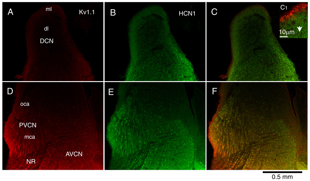
Principal cells of the ventral cochlear nucleus (VCN) differ in the magnitudes of low-voltage-activated potassium (gKL) and hyperpolarization-activated (gh) conductances that determine the time course of signaling. Octopus cells in mice have large gKL (500 nS) and gh (150 nS), bushy cells have smaller gKL (80 nS) and gh (30 nS), and T stellate cells have little gKL and a small gh (20 nS). gKL Arises through potassium channels of which approximately 60% contain Kv1.1 (potassium channels in the shaker or KCNA family) subunits; gh arises through channels that include hyperpolarization and cyclic nucleotide gated (HCN) 1 subunits. The surfaces of cell bodies and dendrites of octopus cells in the dorsocaudal pole, and of similar cells along the ventrolateral edge of the PVCN, were brightly labeled by an antibody against HCN1 that was colocalized with labeling for Kv1.1. More anteriorly neurons with little surface labeling were intermingled among cell bodies and dendrites with surface labeling for both proteins, likely corresponding to T stellate and bushy cells. The membrane-associated labeling patterns for Kv1.1 and HCN1 were consistent with what is known about the distribution and the electrophysiological properties of the principal cells of the VCN. The cytoplasm of large cells and axonal paranodes contained immunofluorescent labeling for only Kv1.1.


Figures

Figure 1
Figure 1
Examples of measurements of gKL (upper traces) and gh (lower traces) in each of the three major types of principal cells are illustrated. Families of voltage pulses were imposed through patch-electrodes in the whole-cell configuration. Because these conductances have overlapping voltage-sensitivity, they were measured in the presence of blockers of other conductances. In all these recordings voltage-sensitive Na+ currents were blocked by 1 µM tetrodotoxin, Ca2+ currents by 0.25 mM Cd2+, and glycinergic and glutamatergic synaptic currents were blocked by 1 µM strychnine and 40 µM DNQX, respectively. In addition gKL was measured in the presence of 50 µM ZD7288, a blocker of gh and gh was measured in the presence of 50 nM α-DTX, a blocker of Kv1 channels.
Figure 2
Figure 2
Labeling for Kv1.1 (left, red) and for HCN1 (middle, green) and the merged image that reveals colocalization in yellow (right) in parasagittal sections of the cochlear nuclear complex show differing patterns. A. The molecular layer (ml) of the DCN revealed brighter staining for Kv1.1 than the deep layer (dl). The labeling was especially bright at the ependymal layer but that is not obvious at the low magnification that is shown. B. The molecular layer was also more brightly stained for HCN1 than the deep layer whereas the granule cell lamina that separates the DCN and VCN shows little staining. C. The difference in labeling between the molecular layer and deep layers is evident in the merged image and is particularly clear at higher magnification in the inset (C1). C1. The ependymal layer is brightly labeled for Kv1.1 whereas the molecular layer shows evenly distributed punctate staining for HCN1. An example of a spot that resembles a paranode is indicated by the arrow. D. The brightest labeling for Kv1.1 was associated with octopus cells in the caudal, posterior VCN. In more medial sections, octopus cells lay in a triangular region in the most caudal and dorsal part of the nucleus but in this relatively lateral section, brightly labeled cells extended ventrally along the posterior edge of the nucleus almost to the nerve root. Labeling for Kv1.1 increased toward the anterior VCN; brightly labeled spots are evident. E. Octopus cells and scattered neurons in the vicinity of the nerve root were brightly labeled for HCN1 among cells that were only weakly labeled. Labeling for HCN1 was bright anteriorly revealing the border between the labeled magnocellular and unlabeled parvocellular regions. The dim labeling of the bundles of auditory nerve fibers contrasts with the brighter staining of the VCN cells. F. The merged image reveals the same patterns as D and E.
Figure 3
Figure 3
Labeling differs between populations of cells. A–C. Within the octopus cell area many cell bodies and processes were brightly labeled for both Kv1.1 and HCN1. Much of the label for Kv1.1 was colocalized with label for HCN1 but some was not. Elongated bright spots (arrows) resemble those associated with myelinated nerve fibers and likely labeled paranodes. D–F. Along the posterior and ventral border of the VCN some cells and processes were brightly labeled for both antigens. The cells and their processes resembled those in the octopus cell area. They were intermingled with cells that were only dimly labeled (<) that occupy the multipolar cell area. G–I. In the nerve root, antibodies against Kv1.1 labeled numerous elongated spots (arrows). The regions near the arrows are shown at higher magnification in Figure 4. Among the bundle of fibers was a single cell in which label for HCN1 was colocalized with label for Kv1.1. J–L. In the AVCN many cell bodies were surrounded with bright label for both antigens but few processes were distinct. Presumably many of these cells were bushy cells whose fine labeled processes could not be resolved. In the AVCN, too, labeling for Kv1.1 revealed bright spots that were presumably labeled paranodes (arrows). All images were from the same section.
Figure 4
Figure 4
Spots labeled for Kv1.1 in the nerve root have structure. Two regions, in the vicinity of each of the arrows in Figure 3G, are shown at higher magnification. Labeled spots are often elongated and seem to comprise pairs of parallel labeled strips.
Figure 5
Figure 5
Comparison of octopus cells within the octopus cell area with brightly labeled cells along the ventral and caudal border of the VCN in different animals. The size of neurons and the diameters of labeled processes were similar, consistent with all being octopus cells. A–C. Brightly labeled neurons near the caudoventral border of the VCN; edge is outside the picture at the left. D–F. Octopus cells in the midst of the octopus cell area. G–H. Brightly labeled neurons near the caudoventral border. As elsewhere in the VCN, elongated spots that are likely to be perinodes of myelinated nerve fibers were labeled for Kv1.1 but not for HCN1 (rightward arrowheads).
Figure 6
Figure 6
Labeling in the AVCN was variable. Cells whose surfaces were strongly labeled lay adjacent to cells whose surfaces were not labeled (example marked with an o). Much of the label for Kv1.1 and for HCN1 was colocalized but as elsewhere in the VCN nodes were labeled brightly with only Kv1.1 (rightward arrowheads).

References

    1. Adamson CL, Reid MA, Mo ZL, Bowne-English J, Davis RL. Firing features and potassium channel content of murine spiral ganglion neurons vary with cochlear location. J Comp Neurol. 2002;447:331–350. - PubMed
    1. Bal R, Oertel D. Hyperpolarization-activated, mixed-cation current (Ih) in octopus cells of the mammalian cochlear nucleus. J Neurophysiol. 2000;84:806–817. - PubMed
    1. Bal R, Oertel D. Potassium currents in octopus cells of the mammalian cochlear nuclei. J Neurophysiol. 2001;86:2299–2311. - PubMed
    1. Barnes-Davies M, Barker MC, Osmani F, Forsythe ID. Kv1 currents mediate a gradient of principal neuron excitability across the tonotopic axis in the rat lateral superior olive. Eur J Neurosci. 2004;19:325–333. - PubMed
    1. Bekele-Arcuri Z, Matos MF, Manganas L, Strassle BW, Monaghan MM, Rhodes KJ, Trimmer JS. Generation and characterization of subtype-specific monoclonal antibodies to K+ channel alpha- and beta-subunit polypeptides. Neuropharm. 1996;35:851–865. - PubMed

Publication types

MeSH terms