. 2008 Jun 13;283(24):16391-9.
doi: 10.1074/jbc.M800945200. Epub 2008 Apr 18.

NF-kappaB-dependent transcriptional activation in lung carcinoma cells by farnesol involves p65/RelA(Ser276) phosphorylation via the MEK-MSK1 signaling pathway

Affiliations

NF-kappaB-dependent transcriptional activation in lung carcinoma cells by farnesol involves p65/RelA(Ser276) phosphorylation via the MEK-MSK1 signaling pathway

Joung Hyuck Joo et al. J Biol Chem. .

Abstract

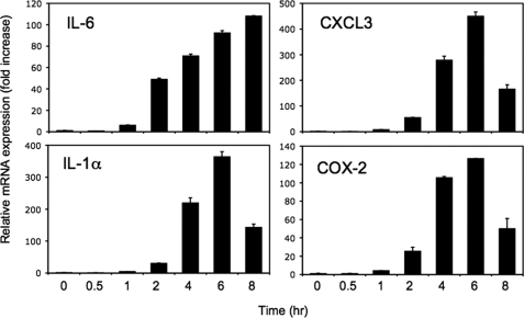
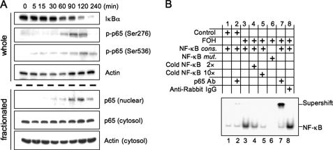
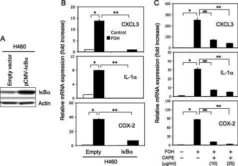
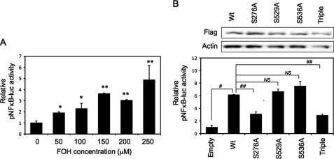
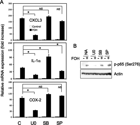
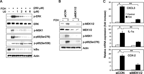
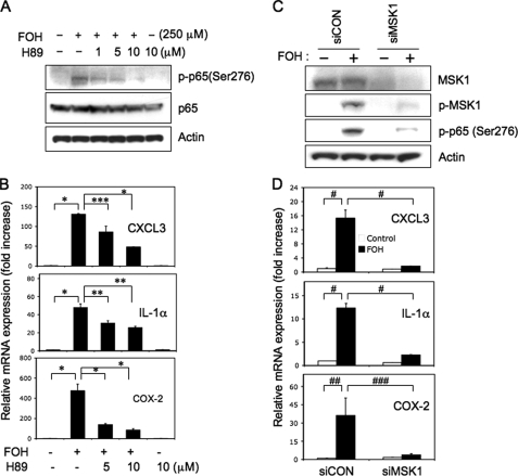
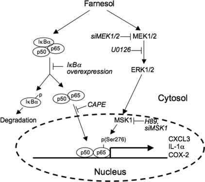
In this study, we demonstrate that treatment of human lung adenocarcinoma H460 cells with farnesol induces the expression of a number of immune response and inflammatory genes, including IL-6, CXCL3, IL-1alpha, and COX-2. This response was dependent on the activation of the NF-kappaB signaling pathway. Farnesol treatment reduces the level of IkappaBalpha and induces translocation of p65/RelA to the nucleus, its phosphorylation at Ser(276), and transactivation of NF-kappaB-dependent transcription. Moreover, overexpression of IkappaBalpha or treatment with the NF-kappaB inhibitor caffeic acid phenethyl ester greatly diminishes the induction of inflammatory gene expression by farnesol. We provide evidence indicating that the farnesol-induced phosphorylation of p65/RelA at Ser(276) is important for optimal transcriptional activity of NF-kappaB. The MEK1/2 inhibitor U0126 and knockdown of MEK1/2 expression with small interfering RNAs effectively blocked the phosphorylation of p65/RelA(Ser(276)) but not that of Ser(536), suggesting that this phosphorylation is dependent on the activation of the MEK1/2-ERK1/2 pathway. We further show that inhibition of MSK1, a kinase acting downstream of MEK1/2-ERK1/2, by H89 or knockdown of MSK1 expression also inhibited phosphorylation of p65/RelA(Ser(276)), suggesting that this phosphorylation is dependent on MSK1. Knockdown of MEK1/2 or MSK1 expression inhibits farnesol-induced expression of CXCL3, IL-1alpha, and COX-2 mRNA. Our results indicate that the induction of inflammatory genes by farnesol is mediated by the activation of the NF-kappaB pathway and involves MEK1/2-ERK1/2-MSK1-dependent phosphorylation of p65/RelA(Ser(276)). The activation of the NF-kappaB pathway by farnesol might be part of a prosurvival response during farnesol-induced ER stress.


Figures

FIGURE 1.
FIGURE 1.
Farnesol induces IL-6, CXCL3, IL-1α, and COX-2 mRNA expression. H460 cells were treated with 250 μm farnesol, and at the times indicated, cells were collected and total RNA was isolated. Levels of IL-6, CXCL3, IL-1α, and COX-2 mRNA expression were evaluated by QRT-PCR as described under “Experimental Procedures.” Each value is the mean ± S.D. of three separate experiments.
FIGURE 2.
FIGURE 2.
Farnesol treatment induces phosphorylation and nuclear localization of p65. A, H460 cells were treated with 250 μm farnesol at the times indicated. Levels of IκBα, phospho-p65(Ser276), and phospho-p65(Ser536) in whole cell lysates were examined by Western blot analysis with the respective antibodies. Levels of p65 were also determined in nuclear and cytosolic fractions. Actin is shown as a control for equal loading. B, EMSA. Nuclear extracts isolated from cells treated for 2 h with vehicle (control) or farnesol (FOH; 250 μm) were incubated with radiolabeled NF-κB consensus oligonucleotide (NF-κB cons.) or NF-κB mutant oligonucleotide (NF-κB mut.) and subjected to EMSA. Antibodies against p65 and anti-rabbit-IgG were used for supershift assay.
FIGURE 3.
FIGURE 3.
Inhibition of the NF-κB signaling pathway reduces the induction of CXCL3, IL-1α, and COX-2 mRNA expression by farnesol. A, analysis of IκBα in H460(IκBα) cells overexpressing IκBα. IκBα levels were examined in H460(Empty) and H460(IκBα) cells by Western blot analysis using an anti-IκBα antibody. Actin is shown as a control for equal loading. B, H460(IκBα) and H460(Empty) cells were treated with 250 μm farnesol (FOH) for 6 h. RNA was then isolated, and the expression of CXCL3, IL-1α, and COX-2 was examined by QRT-PCR. C, H460 cells were pretreated with the NF-κB inhibitor, CAPE, at 10 or 25 μg/ml 2 h before 250 μm farnesol was added. After an additional 6 h of incubation, RNA was harvested and analyzed by QRT-PCR for the expression of CXCL3, IL-1α, and COX-2 mRNA. Each value is the mean ± S.D. of three separate PCRs. *, p < 0.0001; **, p < 0.001.
FIGURE 4.
FIGURE 4.
The Ser276 phosphorylation site in p65-RelA is important in NF-κB-mediated transcriptional activation by farnesol. A, activation of NF-κB-dependent transcriptional activation by farnesol (FOH). H460 cells were transfected with pNF-κB-Luc and subsequently treated with farnesol for 24 h at the concentrations indicated before cells were collected and assayed for luciferase reporter activities. *, p < 0.1; **, p < 0.01 compared with vehicle-treated control (0 μm). B, H460 cells were co-transfected with pNFκB-Luc and expression vectors containing wild type or various point mutants of FLAG-p65-RelA (Empty, empty vector; Wt, FLAG-p65; S276A, FLAG-p65/S276A mutant; S529A, FLAG-p65/S529A; S536A, FLAG-p65/S536A; Triple, FLAG-p65/S276/529/536A triple mutant). After 48 h, cells were treated with 250 μm farnesol, and 24 h later, they were harvested. Cells were assayed for luciferase reporter activities (bottom) or Western blot analysis with anti-FLAG M2 to determine the expression levels of FLAG-p65 proteins (top). Each value is the mean ± S.D. of three separate experiments. #, p < 0.0001; ##, p < 0.001; NS, no significant change.
FIGURE 5.
FIGURE 5.
Optimal induction of CXCL3, IL-1α, and COX-2 mRNA expression by farnesol in H460 cells involves activation of MEK1/2. A, H460 cells were treated for 30 min with vehicle (C), MEK inhibitor U0126 (U0; 5 μm), p38 inhibitor SB203580 (SB; 10 μm), or JNK inhibitor SP600125 (SP; 10 μm) before farnesol (FOH; 250 μm) was added. Cells were collected 6 h later, and the levels of CXCL3, IL-1α, and COX-2 mRNA were analyzed by QRT-PCR. Each value is the mean ± S.D. of three separate PCRs. B, effect of MAPK inhibitors on p65(Ser276) phosphorylation. H460 cells were treated as described for A, except that they were treated for 2 h with farnesol. Protein lysates were then examined by Western blot analysis with an anti-phospho-p65(Ser276) antibody. Actin is shown as a control for equal loading. *, p < 0.01; NS, no significant change.
FIGURE 6.
FIGURE 6.
Farnesol-induced phosphorylation of p65(Ser276) is mediated by the MEK1/2 pathway. A, H460 cells were treated for 30 min with the MEK1/2 inhibitor U0126 (U0) at the concentration indicated before farnesol (FOH; 250 μm) was added. Two h later, total protein lysates were isolated and examined by Western blot analysis using antibodies against phospho-p65(Ser276), phospho-p65(Ser536), phospho-MSK1, IκBα, phospho-ERK, and total ERK (ERK). Actin is shown as a control for equal loading. B, H460 cells were transfected with MEK1/2 (siMEK1/2) or scrambled siRNAs (siCON) and 48 h later treated for 2 h with 250 μm farnesol. Total protein lysates were then prepared and examined by Western blot analysis using antibodies against total MEK1/2 (MEK), phospho-MEK1/2, phospho-MSK1, and phospho-p65(Ser276). Actin is shown as a control for equal loading. C, MEK1/2 or control siRNA-transfected cells were treated with 250 μm farnesol for 6 h before RNA was isolated. Expression of CXCL3, IL-1 α, andCOX-2 mRNA were examined by QRT-PCR. Each value is the mean ± S.D. of three separate PCRs. *, p < 0.0001; **, p < 0.001.
FIGURE 7.
FIGURE 7.
The induction of phosphorylation of p65(Ser276), immune response, and inflammatory genes by farnesol involves activation of MSK1. A, H89 inhibits farnesol-induced phosphorylation of p65(Ser276). H460 cells were treated for 2 h with MSK1 inhibitor H89 before farnesol (FOH) was added. Two h later, protein lysates were examined by Western blot analysis with p-p65(Ser276) and total p65 antibodies. Actin is shown as a control for equal loading. B, H89 inhibits the induction of immune response and inflammatory genes by farnesol. H460 cells were treated H89 as described for A, except that they were treated for 6 h with farnesol before total RNA was isolated. mRNA expression of CXCL3, IL-1α, and COX-2 was then analyzed by QRT-PCR. Each value is the mean ± S.D. of three separate PCRs. *, p < 0.0001; **, p < 0.001; ***, p < 0.01. C, knockdown of MSK1 inhibits farnesol-induced phosphorylation of p65(Ser276). H460 cells were transfected with MSK1 (siMSK1) or scrambled siRNAs (siCON) and 48 h later treated for 2 h with 250 μm farnesol. Total cell lysates were examined by Western blot analysis using antibodies against MSK1, phospho-MSK1, and phospho-p65(Ser276). Actin is shown as a control for equal loading. D, knockdown of MSK1 inhibits the induction of immune response and inflammatory genes by farnesol. H460 cells were transfected with siMSK1 or siCON as described and treated for 6 h with farnesol before total RNA was isolated. CXCL3, IL-1α, and COX-2 mRNA expression was then analyzed by QRT-PCR. Each value is the mean ± S.D. of three separate PCRs. #, p < 0.0001; ##, p < 0.001; ###, p < 0.01.
FIGURE 8.
FIGURE 8.
A schematic model of the role of the NF-κB and MEK1/2 pathways in the induction of inflammatory genes by farnesol. Farnesol treatment reduces the level of IκBα and induces translocation of NF-κB transcription factors to the nucleus. Activation of the MEK1/2-ERK1/2 pathway by farnesol results in the activation of MSK1 and subsequently the phosphorylation of p65 at Ser276 and increased p65 activity. Together this leads to increased CXCL3, IL-1α, and COX-2 expression by NF-κB. Inhibition of MEK1/2 by U0126 or by siRNAs results in reduced MSK1 and p65 phosphorylation, whereas inhibition of MSK1 by H89 or siRNAs represses the phosphorylation of p65 and the induction of these inflammatory genes. (→, induction; ⊢, inhibition).

References

    1. Goldstein, J. L., and Brown, M. S. (1990) Nature 343 425–430 - PubMed
    1. Edwards, P. A., and Ericsson, J. (1999) Annu. Rev. Biochem. 68 157–185 - PubMed
    1. Endres, M., and Laufs, U. (2004) Stroke 35 2708–2711 - PubMed
    1. Hanley, K., Komuves, L. G., Ng, D. C., Schoonjans, K., He, S. S., Lau, P., Bikle, D. D., Williams, M. L., Elias, P. M., Auwerx, J., and Feingold, K. R. (2000) J. Biol. Chem. 275 11484–11491 - PubMed
    1. Rioja, A., Pizzey, A. R., Marson, C. M., and Thomas, N. S. (2000) FEBS Lett. 467 291–295 - PubMed

Publication types

MeSH terms

Associated data