

ApoFnr binds as a monomer to promoters regulating the expression of enterotoxin genes of Bacillus cereus
- PMID: 18424517
- PMCID: PMC2446755
- DOI: 10.1128/JB.00336-08
ApoFnr binds as a monomer to promoters regulating the expression of enterotoxin genes of Bacillus cereus
Abstract
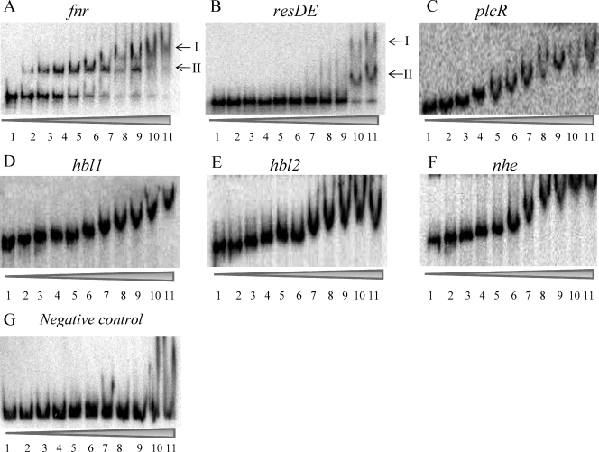
Bacillus cereus Fnr is a member of the Crp/Fnr (cyclic AMP-binding protein/fumarate nitrate reduction regulatory protein) family of helix-turn-helix transcriptional regulators. It is essential for the expression of hbl and nhe enterotoxin genes independently of the oxygen tension in the environment. We studied aerobic Fnr binding to target sites in promoters regulating the expression of enterotoxin genes. B. cereus Fnr was overexpressed and purified as either a C-terminal His-tagged (Fnr(His)) fusion protein or an N-terminal fusion protein tagged with the Strep-tag (IBA BioTAGnology) ((Strep)Fnr). Both recombinant Fnr proteins were produced as apoforms (clusterless) and occurred as mixtures of monomers and oligomers in solution. However, apoFnr(His) was mainly monomeric, while apo(Strep)Fnr was mainly oligomeric, suggesting that the His-tagged C-terminal extremity may interfere with oligomerization. The oligomeric state of apo(Strep)Fnr was dithiothreitol sensitive, underlining the importance of a disulfide bridge for apoFnr oligomerization. Electrophoretic mobility shift assays showed that monomeric apoFnr, but not oligomeric apoFnr, bound to specific sequences located in the promoter regions of the enterotoxin regulators fnr, resDE, and plcR and the structural genes hbl and nhe. The question of whether apoFnr binding is regulated in vivo by redox-dependent oligomerization is discussed.
Figures
Similar articles
-
Bacillus cereus Fnr binds a [4Fe-4S] cluster and forms a ternary complex with ResD and PlcR.BMC Microbiol. 2012 Jun 25;12:125. doi: 10.1186/1471-2180-12-125. BMC Microbiol. 2012. PMID: 22731107 Free PMC article.
-
ResDE-dependent regulation of enterotoxin gene expression in Bacillus cereus: evidence for multiple modes of binding for ResD and interaction with Fnr.J Bacteriol. 2009 Jul;191(13):4419-26. doi: 10.1128/JB.00321-09. Epub 2009 Apr 24. J Bacteriol. 2009. PMID: 19395489 Free PMC article.
-
The redox regulator Fnr is required for fermentative growth and enterotoxin synthesis in Bacillus cereus F4430/73.J Bacteriol. 2007 Apr;189(7):2813-24. doi: 10.1128/JB.01701-06. Epub 2007 Jan 26. J Bacteriol. 2007. PMID: 17259311 Free PMC article.
-
The Food Poisoning Toxins of Bacillus cereus.Toxins (Basel). 2021 Jan 28;13(2):98. doi: 10.3390/toxins13020098. Toxins (Basel). 2021. PMID: 33525722 Free PMC article. Review.
-
FNR and its role in oxygen-regulated gene expression in Escherichia coli.FEMS Microbiol Rev. 1990 Aug;6(4):399-428. doi: 10.1111/j.1574-6968.1990.tb04109.x. FEMS Microbiol Rev. 1990. PMID: 2248796 Review.
Cited by
-
Identification of the dioxygenase-generated intermediate formed during biosynthesis of the dihydropyrrole moiety common to anthramycin and sibiromycin.Bioorg Med Chem. 2015 Feb 1;23(3):449-54. doi: 10.1016/j.bmc.2014.12.024. Epub 2014 Dec 20. Bioorg Med Chem. 2015. PMID: 25564379 Free PMC article.
-
Transcriptional regulation of bacterial virulence gene expression by molecular oxygen and nitric oxide.Virulence. 2014;5(8):794-809. doi: 10.4161/viru.27794. Epub 2014 Oct 31. Virulence. 2014. PMID: 25603427 Free PMC article. Review.
-
The YvfTU two-component system is involved in plcR expression in Bacillus cereus.BMC Microbiol. 2008 Oct 16;8:183. doi: 10.1186/1471-2180-8-183. BMC Microbiol. 2008. PMID: 18925929 Free PMC article.
-
The Role of Fnr Paralogs in Controlling Anaerobic Metabolism in the Diazotroph Paenibacillus polymyxa WLY78.Appl Environ Microbiol. 2020 May 5;86(10):e03012-19. doi: 10.1128/AEM.03012-19. Print 2020 May 5. Appl Environ Microbiol. 2020. PMID: 32198173 Free PMC article.
-
Bacillus cereus Fnr binds a [4Fe-4S] cluster and forms a ternary complex with ResD and PlcR.BMC Microbiol. 2012 Jun 25;12:125. doi: 10.1186/1471-2180-12-125. BMC Microbiol. 2012. PMID: 22731107 Free PMC article.
References
-
- Agaisse, H., M. Gominet, O. A. Okstad, A. B. Kolsto, and D. Lereclus. 1999. PlcR is a pleiotropic regulator of extracellular virulence factor gene expression in Bacillus thuringiensis. Mol. Microbiol. 321043-1053. - PubMed
-
- Busby, S., and R. H. Ebright. 1999. Transcription activation by catabolite activator protein (CAP). J. Mol. Biol. 293199-213. - PubMed
-
- Duport, C., S. Thomassin, G. Bourel, and P. Schmitt. 2004. Anaerobiosis and low specific growth rates enhance hemolysin BL production by Bacillus cereus F4430/73. Arch. Microbiol. 18290-95. - PubMed
-
- Folta-Stogniew, E. 2006. Oligomeric states of proteins determined by size-exclusion chromatography coupled with light scattering, absorbance, and refractive index detectors. Methods Mol. Biol. 32897-112. - PubMed
Publication types
MeSH terms
Substances
LinkOut - more resources
Full Text Sources
Research Materials
Miscellaneous
