. 2008 Jun;190(12):4147-61.
doi: 10.1128/JB.00122-08. Epub 2008 Apr 18.

UpaG, a new member of the trimeric autotransporter family of adhesins in uropathogenic Escherichia coli

Affiliations

UpaG, a new member of the trimeric autotransporter family of adhesins in uropathogenic Escherichia coli

Jaione Valle et al. J Bacteriol. 2008 Jun.

Abstract

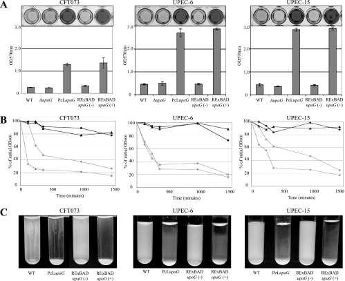
The ability of Escherichia coli to colonize both intestinal and extraintestinal sites is driven by the presence of specific virulence factors, among which are the autotransporter (AT) proteins. Members of the trimeric AT adhesin family are important virulence factors for several gram-negative pathogens and mediate adherence to eukaryotic cells and extracellular matrix (ECM) proteins. In this study, we characterized a new trimeric AT adhesin (UpaG) from uropathogenic E. coli (UPEC). Molecular analysis of UpaG revealed that it is translocated to the cell surface and adopts a multimeric conformation. We demonstrated that UpaG is able to promote cell aggregation and biofilm formation on abiotic surfaces in CFT073 and various UPEC strains. In addition, UpaG expression resulted in the adhesion of CFT073 to human bladder epithelial cells, with specific affinity to fibronectin and laminin. Prevalence analysis revealed that upaG is strongly associated with E. coli strains from the B2 and D phylogenetic groups, while deletion of upaG had no significant effect on the ability of CFT073 to colonize the mouse urinary tract. Thus, UpaG is a novel trimeric AT adhesin from E. coli that mediates aggregation, biofilm formation, and adhesion to various ECM proteins.


Figures

FIG. 1.
FIG. 1.
In silico analysis of the UpaG protein. (A) Schematic illustration of the domain organization of UpaG from E. coli CFT073, YadA from Yersinia enterocolitica, and NhhA from Neisseria meningitidis. Indicated are the signal peptide (S.P.) and the localizations of the Hep-Hag and Him domains (invasin and hemagglutinin domains). Alignments were generated using Clustal W. (B) Multiple-sequence alignment of the translocation units from UpaG, YadA, and NhhA. Identical residues are indicated by shaded boxes, whereas conservative substitutions are indicated by unshaded boxes. The L2, L1, and β subdomains of UpaG are aligned with the corresponding regions from YadA and NhhA. (C) Computer model of the putative 3D structure of UpaG. (a) Monomeric structure; (b) trimeric structure; (c) upper view. The different subregions are in green (β), red (L1), and yellow (L2).
FIG. 2.
FIG. 2.
Surface localization of the UpaG passenger domain. (A) Western blot analysis of UpaG performed using outer membrane fractions from CFT073, CFT073 ΔupaG, CFT073 PcL upaG, and CFT073 RExBAD upaG in the presence (+) or absence (−) of 0.2% arabinose. Outer membrane fractions were resolved by SDS-PAGE in the presence of 8 M urea. The monomeric form of UpaG is indicated (m), as are possible dimers (d) and trimers (t). (B) Immunofluorescence assays of UpaG from CFT073, CFT073 ΔupaG, CFT073 PcL upaG, and CFT073 RExBAD upaG in the presence (+) or absence (−) of 0.2% arabinose. Overnight cultures were fixed and incubated with anti-UpaG serum, followed by incubation with a secondary polyclonal goat anti-rabbit serum coupled to Alexa 488 and DAPI. (C) Immunogold electron microscopy of cell sections of CFT073 and CFT073 PcL upaG probed with anti-UpaG and then with gold-labeled anti-rabbit IgG. Anti-UpaG-labeled gold particles were observed on the surface of CFT073 PcL upaG (but not CFT073) up to a distance of approximately 100 nm (arrows). One CFT073 cell and two individual CFT073 PcL upaG cells are shown as representatives of the average labeling observed for cells in several fields of view.
FIG. 3.
FIG. 3.
Surface localization of the c-Jun leucine zipper in a c-Jun-UpaG chimeric protein. (A) Schematic illustration of recombinant c-Jun-UpaG proteins constructed in this study. The position of the PelB signal sequence (SS), the E-tag, the leucine zipper of c-Jun (noted as Jun), and the L2, L1, and β subdomains are indicated, as is the lac promoter (plac). (B) Immunoblots of whole-cell protein extracts from induced cultures expressing the different chimeric constructions. The blot was probed with anti-E-tag MAb. The UpaGL2-L1-β protein migrated as a polypeptide of 23 kDa despite its calculated mass of 19.8 kDa. The molecular masses of UpaGβ (14.6 kDa) and UpaGL1-β (16.3 kDa) were as calculated. (C) Cell-cell aggregation driven by the interaction of the surface-exposed leucine zipper dimerization domain in the different constructions in the presence (black) or absence (gray) of the inducer IPTG. E. coli UT5600 was used as the control. IPTG-induced and uninduced cultures were left to stand without being shaken. Samples of 100 μl were taken from the top of the cultures (1 cm from the surface) at regular time intervals, and the OD600 was measured. The degree of aggregation is inversely proportional to the turbidity. The OD600 just after the induction was taken as 100%. Data represent the means from three independent experiments. Standard deviations are indicated by error bars. (D) Representative photographs of induced (+) and uninduced (−) E. coli cultures taken after 120 min without shaking.
FIG. 4.
FIG. 4.
Biofilm formation and aggregation phenotypes. (A) Static biofilm formation in polystyrene microtiter plates by CFT073, UPEC-6, and UPEC-15 (and their upaG derivatives) in the presence (+) or absence (−) of 0.2% arabinose, where indicated. Biofilm growth was quantified by the solubilization of crystal violet-stained cells with ethanol-acetone and the determination of the absorbance at 570 nm. WT, wild type. (B) Autoaggregation assay demonstrating the settling profiles from liquid suspensions of CFT073/UPEC-6/UPEC-15 (black circles), CFT073/UPEC-6/UPEC-15 PcL upaG derivatives (gray circles), and CFT073/UPEC-6/UPEC-15 RExBAD upaG derivatives in the presence (gray triangles) or absence (black triangles) of 0.2% arabinose. Cells were diluted to an OD600 of 2.5 in a 3.0-ml volume. One-hundred-microliter samples were taken from the top of the cultures (1 cm from the surface) at regular time intervals, and the OD600 was measured. (C) Photographs of E. coli cultures taken after 24 h without shaking.
FIG. 5.
FIG. 5.
Adhesive properties of UpaG. (A) Adherence of CFT073, CFT073 ΔupaG, and CFT073 PcL upaG strains to T24, HeLa, and BSC1 epithelial cells. Values correspond to the means ± standard deviations from three independent experiments (*, denotes a P of <0.01). WT, wild type. (B) Adherence of CFT073 ΔupaG and CFT073 PcL upaG cells to a mixed monolayer of T24 cells expressing the red fluorescence protein (red cells) and HeLa cells. Actin was labeled by phalloidin Alexa 647 (in blue), revealing all eukaryotic cells. Bacteria were observed using phase-contrast microscopy and colored green using Adobe Illustrator CS software. CFT073 PcL upaG adhered in significantly greater numbers to T24 bladder epithelial cells than to HeLa epithelial cells (left panel). In contrast, CFT073 ΔupaG showed no difference in adherence to T24 bladder epithelial cells and HeLa epithelial cells (right panel).
FIG. 6.
FIG. 6.
Adherence to ECM proteins. Adherence of CFT073, CFT073 ΔupaG, CFT073 PcL upaG, and CFT073 RExBAD upaG in the presence of 0.2% of arabinose (+) or glucose (−) to heparan, laminin, fibronectin, and collagens I, II, and IV. Cells that adhered to ECM proteins were detected with a polyclonal serum raised against E. coli and quantified by ELISA. Data are means (± standard deviations) from two independent experiments. WT, wild type.
FIG. 7.
FIG. 7.
Role of UpaG in virulence. (A) Persistence of CFT073, CFT073 ΔupaG, and CFT073 PcL upaG in the bladders of C57BL/6 mice following intraurethral challenge. At days 1 and 5 following infection, all three strains were recovered in equivalent numbers. (B) Persistence of CFT073, CFT073 ΔupaG, and CFT073 PcL upaG in the bladders and kidneys of C3H/HeJ mice following intraurethral challenge. At day 1 and day 5 following infection, there was no significant difference in the abilities of the three strains to colonize the bladder or kidneys.

Similar articles

Cited by

References

    1. Abate, C., D. Luk, E. Gagne, R. G. Roeder, and T. Curran. 1990. Fos and Jun cooperate in transcriptional regulation via heterologous activation domains. Mol. Cell. Biol. 105532-5535. - PMC - PubMed
    1. Abate, C., D. Luk, R. Gentz, F. J. Rauscher III, and T. Curran. 1990. Expression and purification of the leucine zipper and DNA-binding domains of Fos and Jun: both Fos and Jun contact DNA directly. Proc. Natl. Acad. Sci. USA 871032-1036. - PMC - PubMed
    1. Balligand, G., Y. Laroche, and G. Cornelis. 1985. Genetic analysis of virulence plasmid from a serogroup 9 Yersinia enterocolitica strain: role of outer membrane protein P1 in resistance to human serum and autoagglutination. Infect. Immun. 48782-786. - PMC - PubMed
    1. Barenkamp, S. J. 1996. Immunization with high-molecular-weight adhesion proteins of nontypeable Haemophilus influenzae modifies experimental otitis media in chinchillas. Infect. Immun. 641246-1251. - PMC - PubMed
    1. Belda, E., A. Moya, and F. J. Silva. 2005. Genome rearrangement distances and gene order phylogeny in gamma-Proteobacteria. Mol. Biol. Evol. 221456-1467. - PubMed

Publication types

MeSH terms

LinkOut - more resources