. 2008 Jun;74(11):3434-43.
doi: 10.1128/AEM.02575-07. Epub 2008 Apr 18.

Anaerobic and aerobic degradation of cyanophycin by the denitrifying bacterium Pseudomonas alcaligenes strain DIP1 and role of three other coisolates in a mixed bacterial consortium

Affiliations

Anaerobic and aerobic degradation of cyanophycin by the denitrifying bacterium Pseudomonas alcaligenes strain DIP1 and role of three other coisolates in a mixed bacterial consortium

Ahmed Sallam et al. Appl Environ Microbiol. 2008 Jun.

Abstract

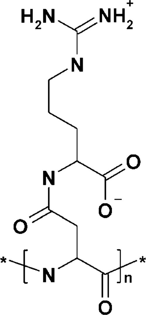
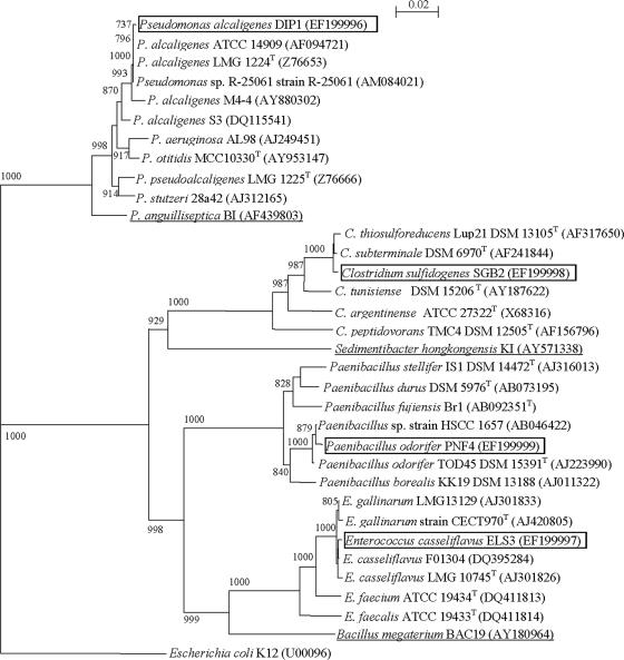
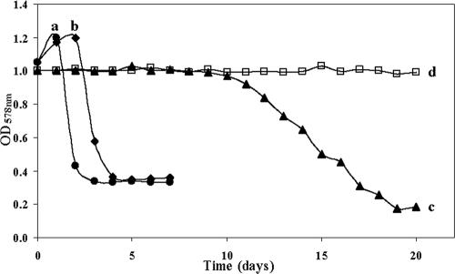
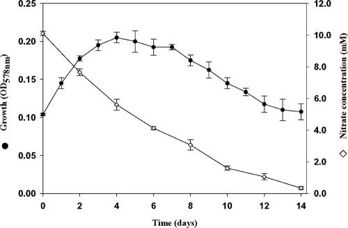
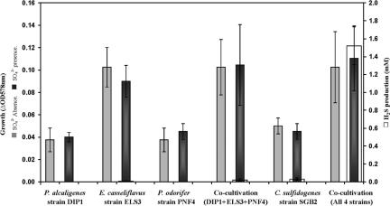
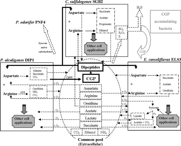
Four bacterial strains were isolated from a cyanophycin granule polypeptide (CGP)-degrading anaerobic consortium, identified by 16S rRNA gene sequencing, and assigned to species of the genera Pseudomonas, Enterococcus, Clostridium, and Paenibacillus. The consortium member responsible for CGP degradation was assigned as Pseudomonas alcaligenes strain DIP1. The growth of and CGP degradation by strain DIP1 under anaerobic conditions were enhanced but not dependent on the presence of nitrate as an electron acceptor. CGP was hydrolyzed to its constituting beta-Asp-Arg dipeptides, which were then completely utilized within 25 and 4 days under anaerobic and aerobic conditions, respectively. The end products of CGP degradation by strain DIP1 were alanine, succinate, and ornithine as determined by high-performance liquid chromatography analysis. The facultative anaerobic Enterococcus casseliflavus strain ELS3 and the strictly anaerobic Clostridium sulfidogenes strain SGB2 were coisolates and utilized the beta-linked isodipeptides from the common pool available to the mixed consortium, while the fourth isolate, Paenibacillus odorifer strain PNF4, did not play a direct role in the biodegradation of CGP. Several syntrophic interactions affecting CGP degradation, such as substrate utilization, the reduction of electron acceptors, and aeration, were elucidated. This study demonstrates the first investigation of CGP degradation under both anaerobic and aerobic conditions by one bacterial strain, with regard to the physiological role of other bacteria in a mixed consortium.


Figures

FIG. 1.
FIG. 1.
Chemical structure of the aspartic acid-arginine building block of CGP (34).
FIG. 2.
FIG. 2.
Degradation of CGP in liquid and solid media. (A) Four anaerobic Hungate tubes, which contained initial concentrations of 1 g liter−1 CGP plus 0.5 g liter−1 yeast extract in 10 ml BM, after incubation at 30°C for 15 days. Tube 1 contains P. alcaligenes strain DIP1 and shows CGP degradation after the first 48 h of incubation. Tubes 2, 3, and 4 contain E. casseliflavus strain ELS3, C. sulfidogenes strain SGB2, and P. odorifer strain PNF4, respectively. None of the three strains could degrade CGP. (B) Degradation halo by P. alcaligenes strain DIP1 appearing after 24 h of incubation on an aerobic CGP overlay agar plate. (C) Degradation halo by P. alcaligenes strain DIP1 appearing after 3 to 4 days of incubation on an anaerobic CGP overlay agar plate.
FIG. 3.
FIG. 3.
Neighbor-joining tree based on 16S rRNA gene sequences showing the estimated phylogenetic relationships of the four consortium members to their closely related species and other bacteria. The underlined strains were previously investigated for CGP degradation (30, 31, 33). The names of species investigated in this study are boxed in the diagram. E. coli K-12 was used as an out-group. Accession numbers are given in parentheses. Bootstrap values are shown as percentages of 1,000 replicates. Scale bar = 2% sequence divergence.
FIG. 4.
FIG. 4.
Degradation of 1 g liter−1 CGP in 10 ml BM by axenic cultures of P. alcaligenes strain DIP1 in Hungate tubes cultivated anaerobically at 30°C under shaking conditions. a, tube containing 0.5 g liter−1 yeast extract and 10 mM sodium nitrate and showing the fastest CGP degradation by P. alcaligenes strain DIP1 with visual disappearance of CGP and the lowest OD578 nm after one and four incubation days, respectively; b, tube containing 0.5 g liter−1 yeast extract and no sodium nitrate and showing the visual disappearance of CGP and the lowest OD578 nm after four and five incubation days, respectively; c, tube containing 10 mM sodium nitrate and no yeast extract and showing the visual disappearance of CGP and the lowest OD578 nm after 12 and 19 incubation days, respectively; d, tube containing neither nitrate nor yeast extract (negative control).
FIG. 5.
FIG. 5.
Growth of and nitrate reduction by P. alcaligenes DIP1 when grown at 30°C under anaerobic conditions. Duplicate Hungate tubes containing 10 ml BM with 0.5% (wt/vol) Casamino Acids and 10 mM sodium nitrate were used. Growth (measured by the OD578 nm) and the concentration of nitrate in the medium during the incubation period are shown.
FIG. 6.
FIG. 6.
Growth and H2S production by the consortium and individually by the four isolates. Cells were incubated at 30°C in duplicate BM tubes containing 0.5% (wt/vol) Casamino Acids with or without 20 mM sodium sulfate as an electron acceptor. C. sulfidogenes strain SGB2 produced 1.5 mM H2S only when cocultivated with the other three consortium members. Axenic cultures of the four members or other combinations thereof did not produce significant amounts of H2S.
FIG. 7.
FIG. 7.
Ecological and physiological interactions between the four members of the consortium. Various aspects of contribution between P. alcaligenes DIP1, E. casseliflavus ELS3, C. sulfidogenes SGB2, and P. odorifer PNF4 are shown with CGP degradation in the center. See the text for a detailed description. O2, only under an aerobic atmosphere; ±O2, regardless of the state of aeration. The dotted fields represent hypothetical aspects.

References

    1. Aboulmagd, E., F. B. Oppermann-Sanio, and A. Steinbüchel. 2000. Molecular characterization of the cyanophycin synthetase from Synechocystis sp. strain PCC6308. Arch. Microbiol. 174:297-306. - PubMed
    1. Achouak, W., P. Normand, and T. Heulin. 1999. Comparative phylogeny of rrs and nifH genes in the Bacillaceae. Int. J. Syst. Bacteriol. 49:961-967. - PubMed
    1. Allen, M. M., and M. A. Hawley. 1983. Protein degradation and synthesis of cyanophycin granule polypeptide in Aphanocapsa sp. J. Bacteriol. 154:1480-1484. - PMC - PubMed
    1. Appanna, V. D., R. Hamel, C. Mackenzie, P. Kumar, and S. Kalyuzhnyi. 2003. Adaptation of Pseudomonas fluorescens to Al-citrate: involvement of tricarboxylic acid and glyoxylate cycle enzymes and the influence of phosphate. Curr. Microbiol. 47:521-527. - PubMed
    1. Ariño, X., J. J. Ortega-Calvo, M. Hernandez-Marine, and C. Saiz-Jimenez. 1995. Effect of sulfur starvation on the morphology and ultrastructure of the cyanobacterium Gloeothece sp. PCC 6909. Arch. Microbiol. 163:447-453.

MeSH terms

Associated data

LinkOut - more resources