. 2008 May 1;180(9):5794-8.
doi: 10.4049/jimmunol.180.9.5794.

Cutting edge: Foxp3-mediated induction of pim 2 allows human T regulatory cells to preferentially expand in rapamycin

Affiliations

Cutting edge: Foxp3-mediated induction of pim 2 allows human T regulatory cells to preferentially expand in rapamycin

Samik Basu et al. J Immunol. .

Abstract

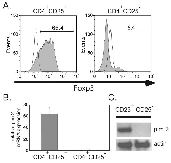
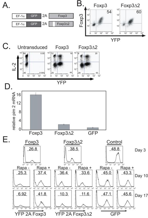
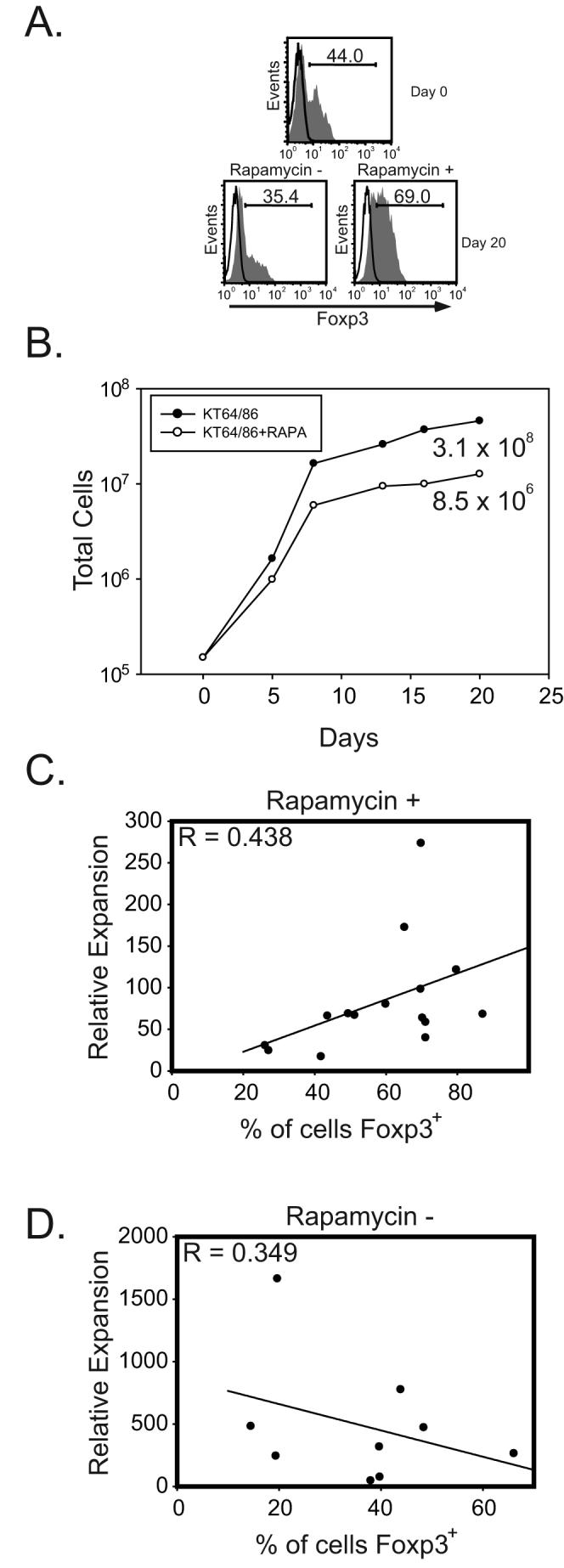
Addition of rapamycin to cultures of expanding natural CD4+CD25+Foxp3+ T regulatory cells (Tregs) helps maintain their suppressive activity, but the underlying mechanism is unclear. Pim 2 is a serine/threonine kinase that can confer rapamycin resistance. Unexpectedly, pim 2 was found to be constitutively expressed in freshly isolated, resting Tregs, but not in CD4+CD25- T effector cells. Introduction of Foxp3, but not Foxp3Delta2, into effector T cells induced pim 2 expression and conferred preferential expansion in the presence of rapamycin, indicating that Foxp3 can regulate pim 2 expression. Finally, we determined there is a positive correlation between Treg expansion and Foxp3 expression in the presence of rapamycin. Together, these results indicate that Tregs are programmed to be resistant to rapamycin, providing further rationale for why this immunosuppressive drug should be used in conjunction with expanded Tregs.


Figures

Figure 1
Figure 1. Natural, Foxp3 expressing Tregs Constitutively Express Pim 2
A. Diagram of Foxp3 expression vectorsFreshly purified CD4+CD25+ Tregs (left panel) and CD4+CD25 T effector cells (right panel) were stained with anti-Foxp3 Ab (shaded histograms) or an isotype control Ab (open histograms) and analyzed by flow cytometry. B. 28S RNA-normalized mRNA levels of pim 2 were measured from freshly isolated CD4+CD25+ and CD4+CD25 T lymphocyte populations as determined by quantitative RT-PCR analysis. The data plotted as the mean ± SD of triplicate determinations from an individual donor. C. Western blot detection of pim 2 and actin (loading control) from freshly isolated CD4+CD25+ and CD4+CD25 T cell populations shown in A. Lysates correspond to 5 × 10⁵ cell equivalents. Data is representative of 3 independent experiments.
Figure 2
Figure 2. Foxp3, but not Foxp3Δ2 Expression, Induces Pim 2 and Confers Rapamycin Resistance
A. Diagram of constructs used in this study. B. CD4+CD25 T cells were activated with CD3/CD28 Ab coated beads and transduced with lentiviral vectors expressing either YFP-2A-Foxp3 or YFP-2A-Foxp3Δ2. The correlation between Foxp3 and YFP expression was determined by flow cytometry. C. YFP-2A-Foxp3, YFP-2A-Foxp3Δ2 and untransduced CD4+CD25 T cells were re-stimulated with PMA (50 ng/ml) and ionomycin (500 ng/ml) for 4 hours and intracellular IL-2 expression was detected by flow cytometry. D. 28S-normalized mRNA levels of pim 2 from either YFP-2A-Foxp3, YFP-2A-Foxp3Δ2, or GFP transduced CD4+CD25 T cells shown in A. was determined by quantitative RT-PCR analysis. The cells were harvested 17 days after transduction. The data are plotted as the mean ± SD of triplicate determinations from one donor. E. CD4+CD25 T cells were activated with CD3/CD28 Ab coated beads and transduced with lentiviral vectors expressing YFP-2A-Foxp3, YFP-2A-Foxp3Δ2 or GFP. 3 days post-transduction the percentage of transduced cells was determined and half the culture was placed in rapamycin (100 ng/ml) containing medium. After an additional 7 (middle panels) or 14 (bottom panels) days of culture, the percentage of transduced cells was measured by flow cytometry. Data is representative of 3 independent experiments.
Figure 3
Figure 3. Natural T regulatory cell expansion in the presence of rapamycin correlates with Foxp3 expression
A. Tregs were enriched by CD25 selection and stained for Foxp3 pre- and post-expansion with irradiated, anti-CD3 Ab loaded KT64 CD86 aAPCs. B. Overall relative expansion of T cells from day 0 to day 20 in the presence or absence of rapamycin (100ng/ml). A. and B. are from the same experiment and are representative of data collected using 9 different donors. C. Enriched Tregs were expanded with rapamycin (100 ng/ml) and the percentage of Foxp3 expressing cells at the end of culture was determined by flow cytometry. Data are compiled from 15 independent experiments. D. Enriched Tregs were expanded without rapamycin and the percentage of Foxp3 expressing cells at the end of culture was determined by flow cytometry. Data are compiled from 9 independent experiments. Correlation between Foxp3 expression and relative cell expansion was determined by nonlinear regression analysis.

References

    1. Bluestone JA. Regulatory T-cell therapy: is it ready for the clinic? Nat Rev Immunol. 2005;5:343–349. - PubMed
    1. Roncarolo MG, Battaglia M. Regulatory T-cell immunotherapy for tolerance to self antigens and alloantigens in humans. Nat Rev Immunol. 2007;7:585–598. - PubMed
    1. Battaglia M, Stabilini A, Roncarolo MG. Rapamycin selectively expands CD4+CD25+FoxP3+ regulatory T cells. Blood. 2005;105:4743–4748. - PubMed
    1. Battaglia M, Stabilini A, Migliavacca B, Horejs-Hoeck J, Kaupper T, Roncarolo MG. Rapamycin promotes expansion of functional CD4+CD25+FOXP3+ regulatory T cells of both healthy subjects and type 1 diabetic patients. J Immunol. 2006;177:8338–8347. - PubMed
    1. Coenen JJA, Koenen HJPM, van Rijssen E, Hilbrands LB, Joosten I. Rapamycin, and not cyclosporin A, preserves the highly suppressive CD27+ subset of human CD4+CD25+ regulatory T cells. Blood. 2006;107:1018–1023. - PubMed

Publication types

MeSH terms