. 2008 Aug;49(8):1715-24.
doi: 10.1194/jlr.M700580-JLR200. Epub 2008 Apr 19.

Xanthophylls are preferentially taken up compared with beta-carotene by retinal cells via a SRBI-dependent mechanism

Affiliations

Xanthophylls are preferentially taken up compared with beta-carotene by retinal cells via a SRBI-dependent mechanism

Alexandrine During et al. J Lipid Res. 2008 Aug.

Abstract

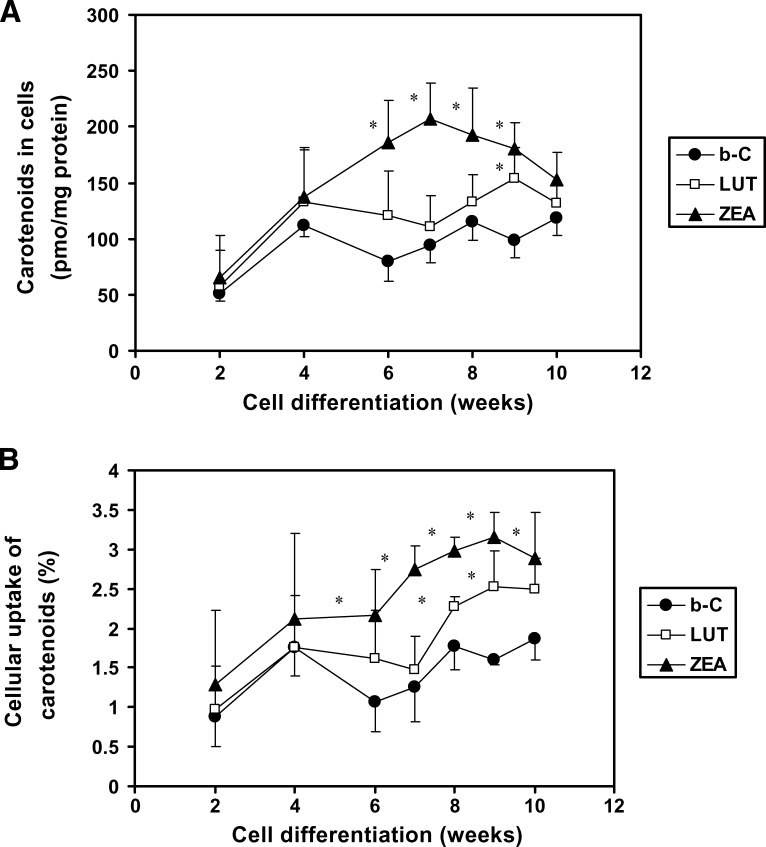
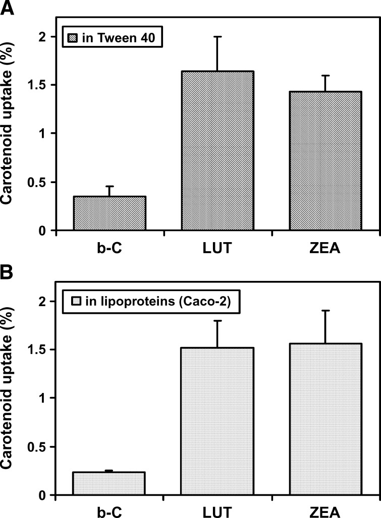
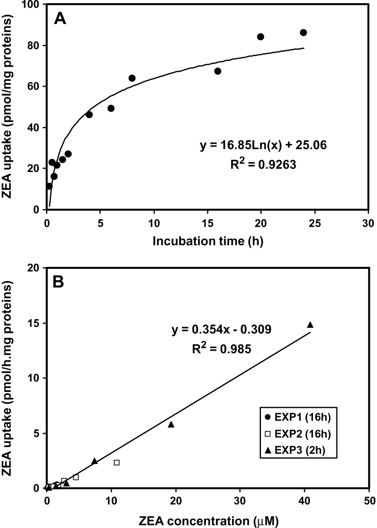
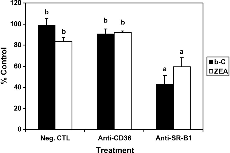
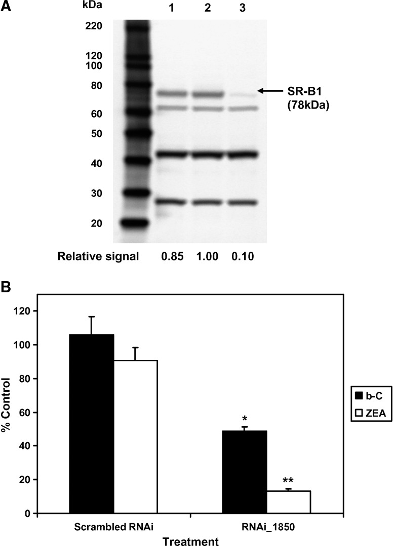
The purpose of this study was to investigate the mechanisms by which carotenoids [xanthophylls vs. beta-carotene(beta-C)] are taken up by retinal pigment epithelial (RPE) cells. The human RPE cell line, ARPE-19, was used. When ARPE-19 cells were fully differentiated (7-9 weeks), the xanthophylls lutein (LUT) and zeaxanthin (ZEA) were taken up by cells to an extent 2-fold higher than beta-C (P < 0.05). At 9 weeks, cellular uptakes were 1.6, 2.5, and 3.2%, respectively, for beta-C, LUT, and ZEA. Similar extents were observed when carotenoids were delivered in either Tween 40 or "chylomicrons" produced by Caco-2 cells. Differentiated ARPE-19 cells did not exhibit any detectable beta-C 15,15'-oxygenase activity or convert exogenous beta-C into vitamin A. When using specific antibodies against the lipid transporters cluster determinant 36 (CD36) and scavenger receptor class B type I (SR-BI), cellular uptake of beta-C and ZEA were significantly decreased (40-60%) with anti-SR-BI but not with anti-CD36. Small interfering RNA transfection for SR-BI led to marked knockdown of SR-BI protein expression (approximately 90%), which resulted in decreased beta-C and ZEA uptakes by 51% and 87%, respectively. Thus, the present data show that RPE cells preferentially take up xanthophylls versus the carotene by a process that appears to be entirely SR-BI-dependent for ZEA and partly so for beta-C. This mechanism may explain, in part, the preferential accumulation of xanthophylls in the macula of the retina.


Figures

Fig. 1.
Fig. 1.
HPLC profiles of a standard lutein (LUT) and zeaxanthin (ZEA) mixture (A), an extract from 7 week differentiated ARPE-19 cells after 20 h of incubation with 1 μM LUT (B), and an extract from 7 week differentiated ARPE-19 cells after 20 h of incubation with 1 μM ZEA (C). HPLC conditions were as follows: a YMC C30 column (250 × 4.6 mm, 3 μm), methanol-methyl-tert-butyl-ether (90:10, v/v) as mobile phase, and a flow rate at 0.9 ml/min (see Materials and Methods). A, B, and C, unidentified peaks; AU, absorbance units; PS, peak solvent.
Fig. 2.
Fig. 2.
Effects of the differentiation stage of ARPE-19 cells on the cellular uptake of β-carotene [β-C (b-C)], LUT, and ZEA. Cells were plated on six-well plates to reach confluence after 7 days, the starting point of the experiment. At different points of differentiation (up to 10 weeks), cells were incubated with β-C, LUT, or ZEA at 1 μM for 18 h. After incubation, cells were analyzed for their carotenoid content. Values are expressed as picomoles of carotenoid per milligram of protein (A) or as percentage of carotenoid recovered in cells from the initial amount added to the cell culture media (B). Data are means ± SD of three independent experiments. * P < 0.05 compared with β-C at a given time point.
Fig. 3.
Fig. 3.
Effects of the mode of delivery of carotenoids to ARPE-19 cells on the cellular uptake of β-C (b-C), LUT, and ZEA. A: β-C, LUT, or ZEA at 1 μM was solubilized in Tween 40 as described previously (32) and detailed in Materials and Methods. The resulting carotenoid-Tween suspension was applied to 7 week differentiated ARPE-19 cells. B: At time 0 of the experiment, 3 week differentiated Caco-2 cell monolayers on inserts were placed on top of 7 week differentiated ARPE-19 cells, and β-C, LUT, or ZEA at 10 μM in Tween 40 was applied at the apical side of Caco-2 cells in the presence of oleic acid and taurocholate to produce carotenoid-enriched chylomicrons (31). Carotenoids were thus delivered to ARPE-19 cells via lipoproteins produced by Caco-2 cells at a final concentration of 1 μM. After 18 h of exposure with one of the carotenoids (either solubilized in Tween 40 or carried by lipoproteins), carotenoid contents in cells and cell culture media were determined by HPLC. Data are means ± SD of two independent experiments.
Fig. 4.
Fig. 4.
Kinetics of ZEA uptake in differentiated ARPE-19 cells as a function of the incubation time (A) and the initial concentration of ZEA (B). At 7 to 8 weeks of differentiation, cells were incubated with ZEA at 1 μM for varying times (15 min up to 24 h) or with varying concentrations of ZEA (0.37 up to 40 μM) for 2 or 16 h. After incubation, cells were analyzed for their ZEA content by HPLC analysis. Data are duplicates from three independent experiments.
Fig. 5.
Fig. 5.
Effects of antibodies against scavenger receptor class B type I (SR-BI; anti-SR-BI) and cluster determinant 36 (CD36; anti-CD36) on β-C (b-C) and ZEA uptake by ARPE-19 cells. At 7 weeks of differentiation, cells were preincubated with nonimmunized rabbit anti-IgG, anti-CD36, or anti-SR-BI at 8 μg protein/ml for 2 h, followed by incubation with β-C or ZEA in Tween 40 for 18 h. The absolute control experiment corresponded to cells incubated with the carotenoid alone (without pretreatment). The negative control (Neg. CTL) corresponded to the nonimmunized rabbit anti-IgG pretreatment. After incubation with carotenoids, cells were harvested and their carotenoid contents analyzed by HPLC. Data are expressed as percentage of the absolute control and presented as means ± SD of three independent experiments. a,b P < 0.001.
Fig. 6.
Fig. 6.
Effects of the small interfering RNA inhibition of SR-BI expression on the cellular uptake of β-C (b-C) and ZEA in ARPE-19 cells. A: Immunoblots of SR-BI expression (using 25 μg total protein/well) in cells treated under the following conditions: lane 1, scrambled RNA interference (RNAi); lane 2, Lipofectamine 2000 (LP2000) only; lane 3, RNAi_1850. The first lane represents protein standards (MagicMark XP Standard from 60 to 220 kDa). B: Cellular uptake of carotenoids (expressed as percentage of control cells treated with LP2000 only) after incubation of 7 week differentiated cells with a carotenoid at 2 μM for 1 h at 72 h after transfection with a RNAi against SR-BI. Data are means ± SD of three independent experiments for each compound tested. * P < 0.005, ** P < 0.0001 compared with the negative control.

References

    1. Moeller S. M., N. Parekh, L. Tinker, C. Ritenbaugh, B. Blodi, R. B. Wallace, and J. A. Mares. 2006. Associations between intermediate age-related macular degeneration and lutein and zeaxanthin in the Carotenoids in Age-Related Eye Disease Study (CAREDS): ancillary study of the Women's Health Initiative. Arch. Ophthalmol. 124 1151–1162. - PubMed
    1. Snodderly D. M. 1995. Evidence for protection against age-related macular degeneration by carotenoids and antioxidant vitamins. Am. J. Clin. Nutr. 62 (6Suppl.) 1448S–1461S. - PubMed
    1. Mozaffarieh M., S. Sacu, and A. Wedrich. 2003. The role of the carotenoids, lutein and zeaxanthin, in protecting against age-related macular degeneration: a review based on controversial evidence. Nutr. J. 11 20–28. - PMC - PubMed
    1. Parker R. S. 1989. Carotenoids in human blood and tissues. J. Nutr. 119 101–104. - PubMed
    1. Goodwin, T. W., and G. Britton. 1988. Distribution and analysis of carotenoids. In Plant Pigments, T.W. Goodwin, editor. Academic Press, New York. 61–127.

Publication types