

Collagens, proteoglycans, MMP-2, MMP-9 and TIMPs in human achilles tendon rupture
- PMID: 18425559
- PMCID: PMC2505242
- DOI: 10.1007/s11999-008-0255-y
Collagens, proteoglycans, MMP-2, MMP-9 and TIMPs in human achilles tendon rupture
Abstract
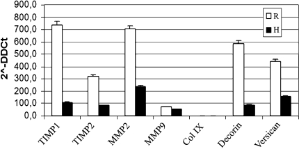
Tendon integrity depends on the extracellular matrix (ECM) metabolism which is regulated by proteolytic enzymes. However, it is unclear which enzymes play a role in tendon rupture. We studied the ECM of 19 ruptured human Achilles tendons, comparing the composition of specimens harvested close to the rupture with specimens harvested from an apparently healthy area in the same tendon. We compared gene expression of collagen Type I, decorin, and versican including enzymes involved in their metabolism as matrix metalloproteases (MMP-2 and -9) and tissue inhibitory of metalloproteinase (TIMP-1 and -2) using real-time PCR, zymography and FACE analysis. We found greater gene expression of proteoglycan core protein decorin and versican, collagen Type I, MMPs and TIMPs in the tendon rupture. Zymography analysis, reflecting expression of enzymatic activity, confirmed the gene expression data at protein level. Carbohydrate content was greater in the macroscopically healthy area than in the ruptured area. In the ruptured area, we found increased core protein synthesis but without the normal glycosaminoglycan production. The tissue in the area of rupture undergoes marked rearrangement at molecular levels and supports the role of MMPs in the pathology.
Figures


References
-
- {'text': '', 'ref_index': 1, 'ids': [{'type': 'DOI', 'value': '10.1016/S0736-0266(03)00107-4', 'is_inner': False, 'url': 'https://doi.org/10.1016/s0736-0266(03)00107-4'}, {'type': 'PubMed', 'value': '14554207', 'is_inner': True, 'url': 'https://pubmed.ncbi.nlm.nih.gov/14554207/'}]}
- Alfredson H, Lorentzon M, Backman S, Backman A, Lerner UH. cDNA-arrays and real-time quantitative PCR techniques in the investigation of chronic Achilles tendinosis. J Orthop Res. 2003;21:970–975. - PubMed
-
- {'text': '', 'ref_index': 1, 'ids': [{'type': 'PubMed', 'value': '13660717', 'is_inner': True, 'url': 'https://pubmed.ncbi.nlm.nih.gov/13660717/'}]}
- Arner O, Lindholm A, Orell SR. Histologic changes in subcutaneous rupture of the Achilles tendon; a study of 74 cases. Acta Chir Scand. 1959;116:484–490. - PubMed
-
- {'text': '', 'ref_index': 1, 'ids': [{'type': 'DOI', 'value': '10.1177/0363546506296043', 'is_inner': False, 'url': 'https://doi.org/10.1177/0363546506296043'}, {'type': 'PubMed', 'value': '17293464', 'is_inner': True, 'url': 'https://pubmed.ncbi.nlm.nih.gov/17293464/'}]}
- Arnoczky SP, Lavagnino M, Egerbacher M, Caballero O, Gardner K. Matrix metalloproteinase inhibitors prevent a decrease in the mechanical properties of stress-deprived tendons: an in vitro experimental study. Am J Sports Med. 2007;35:763–769. - PubMed
-
- {'text': '', 'ref_index': 1, 'ids': [{'type': 'DOI', 'value': '10.1093/glycob/10.3.283', 'is_inner': False, 'url': 'https://doi.org/10.1093/glycob/10.3.283'}, {'type': 'PubMed', 'value': '10704527', 'is_inner': True, 'url': 'https://pubmed.ncbi.nlm.nih.gov/10704527/'}]}
- Calabro A, Hascall VC, Midura RJ. Adaptation of FACE methodology for microanalysis of total hyaluronan and chondroitin sulfate composition from cartilage. Glycobiology. 2000;10:283–293. - PubMed
-
- {'text': '', 'ref_index': 1, 'ids': [{'type': 'DOI', 'value': '10.1016/S0736-0266(02)00016-5', 'is_inner': False, 'url': 'https://doi.org/10.1016/s0736-0266(02)00016-5'}, {'type': 'PubMed', 'value': '12382955', 'is_inner': True, 'url': 'https://pubmed.ncbi.nlm.nih.gov/12382955/'}]}
- Choi HR, Kondo S, Hirose K, Ishiguro N, Hasegawa Y, Iwata H. Expression and enzymatic activity of MMP-2 during healing process of the acute supraspinatus tendon tear in rabbits. J Orthop Res. 2002;20:927–933. - PubMed
MeSH terms
Substances
LinkOut - more resources
Full Text Sources
Research Materials
Miscellaneous
