

Spatiotemporal protein distribution of TGF-betas, their receptors, and extracellular matrix molecules during embryonic tendon development
- PMID: 18425852
- PMCID: PMC3612428
- DOI: 10.1002/dvdy.21547
Spatiotemporal protein distribution of TGF-betas, their receptors, and extracellular matrix molecules during embryonic tendon development
Abstract
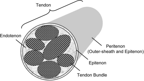
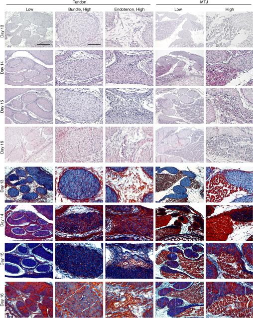
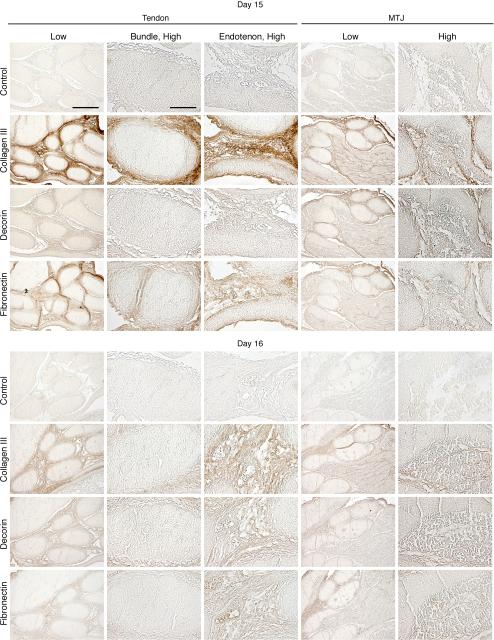
Tendon is one of the least understood tissues of the musculoskeletal system in terms of development and morphogenesis. Collagen fibrillogenesis has been the most studied aspect of tendon development, focusing largely on the role of matrix molecules such as collagen type III and decorin. While involvement of matrix molecules in collagen fibrillogenesis during chick tendon development is well understood, the role of growth factors has yet to be elucidated. This work examines the expression patterns of transforming growth factor (TGF) -beta1, -beta2, and -beta3, and their receptors with respect to expression patterns of collagen type III, decorin, and fibronectin. We focus on the intermediate stages of tendon development in the chick embryo, a period during which the tendon micro- and macro-architecture are being established. Our findings demonstrate for the first time that TGF-beta1, -beta2, and -beta3 have distinct spatiotemporal developmental protein localization patterns in the developing tendon and strongly suggest that these isoforms have independent roles in tendon development.
Figures


References
-
- Anaguchi Y, Yasuda K, Majima T, Tohyama H, Minami A, Hayashi K. The effect of transforming growth factor-beta on mechanical properties of the fibrous tissue regenerated in the patellar tendon after resecting the central portion. Clin Biomech (Bristol, Avon) 2005;20:959–965. - PubMed
-
- Balza E, Borsi L, Allemanni G, Zardi L. Transforming growth factor beta regulates the levels of different fibronectin isoforms in normal human cultured fibroblasts. FEBS Lett. 1988;228:42–44. - PubMed
-
- Birk DE, Mayne R. Localization of collagen types I, III and V during tendon development. Changes in collagen types I and III are correlated with changes in fibril diameter. Eur J Cell Biol. 1997;72:352–361. - PubMed
-
- Birk DE, Nurminskaya MV, Zycband EI. Collagen fibrillogenesis in situ: fibril segments undergo post-depositional modifications resulting in linear and lateral growth during matrix development. Dev Dyn. 1995;202:229–243. - PubMed
-
- Birk DE, Southern JF, Zycband EI, Fallon JT, Trelstad RL. Collagen fibril bundles: a branching assembly unit in tendon morphogenesis. Development. 1989;107:437–443. - PubMed
Publication types
MeSH terms
Substances
Grants and funding
LinkOut - more resources
Full Text Sources
Molecular Biology Databases
Miscellaneous
