. 2008 Jul;76(7):2812-21.
doi: 10.1128/IAI.00126-08. Epub 2008 Apr 21.

Role of aromatic amino acids in receptor binding activity and subunit assembly of the cytolethal distending toxin of Aggregatibacter actinomycetemcomitans

Affiliations

Role of aromatic amino acids in receptor binding activity and subunit assembly of the cytolethal distending toxin of Aggregatibacter actinomycetemcomitans

Linsen Cao et al. Infect Immun. 2008 Jul.

Abstract

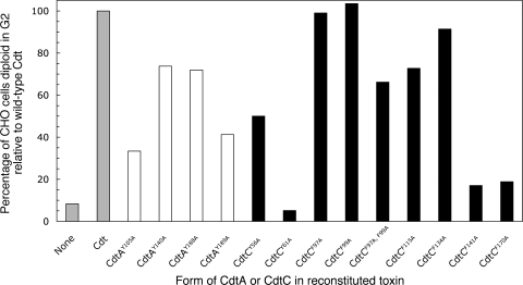
The periodontal pathogen Aggregatibacter actinomycetemcomitans produces a cytolethal distending toxin (Cdt) that inhibits the proliferation of oral epithelial cells. Structural models suggest that the CdtA and CdtC subunits of the Cdt heterotrimer form two putative lectin domains with a central groove. A region of CdtA rich in heterocyclic amino acids (aromatic patch) appears to play an important role in receptor recognition. In this study site-specific mutagenesis was used to assess the contributions of aromatic amino acids (tyrosine and phenylalanine) to receptor binding and CdtA-CdtC assembly. Predominant surface-exposed aromatic residues that are adjacent to the aromatic patch region in CdtA or are near the groove located at the junction of CdtA and CdtC were studied. Separately replacing residues Y105, Y140, Y188, and Y189 with alanine in CdtA resulted in differential effects on binding related to residue position within the aromatic region. The data indicate that an extensive receptor binding domain extends from the groove across the entire face of CdtA that is oriented 180 degrees from the CdtB subunit. Replacement of residue Y105 in CdtA and residues Y61 and F141 in CdtC, which are located in or at the periphery of the groove, inhibited toxin assembly. Taken together, these results, along with the lack of an aromatic amino acid-rich region in CdtC similar to that in CdtA, suggest that binding of the heterotoxin to its cell surface receptor is mediated predominantly by the CdtA subunit. These findings are important for developing strategies designed to block the activity of this prominent virulence factor.


Figures

FIG. 1.
FIG. 1.
Positions of the substitutions for surface-exposed aromatic amino acids in CdtA and CdtC: alignment of the deduced amino acid sequences of A. actinomycetemcomitans recombinant CdtA-His6 and CdtC-His6. The underlined substitution is not a surface-exposed residue. Identical residues are indicated by shading. Predicted signal sequence cleavage sites and amino termini of the proteins resolved in the crystal structure are indicated by the small and large arrows, respectively. Predicted disulfides are indicated by connecting lines.
FIG. 2.
FIG. 2.
Effects of single aromatic amino acid substitutions on the binding of CdtA-His6 and CdtC-His6 to CHO cells. (A) Wild-type or mutated CdtA-His6 proteins (10 μg/well) were added to wells containing 1.5 × 10⁴ cells. Bound protein was detected with anti-His·Tag monoclonal antibody (1:3,000 dilution) and anti-mouse IgG-horseradish peroxidase conjugate (1:3,000 dilution). (B) Wild-type CdtA and CdtA(Y188A) were incubated in reconstitution buffer alone and with CdtC or with CdtB plus CdtC. The preparations (4 μg of CdtA) were added to wells containing CHO cells as described above for panel A. Bound protein was detected as described above, and absorbance ratios were calculated relative to the binding of CdtA or CdtA(Y188A). (C) Wild-type or mutated CdtC-His6 proteins run under the same conditions that were used in the experiment whose results are shown in panel A. All samples were run in triplicate. Statistically significant differences between the mutated and wild-type proteins are indicated by asterisks (one asterisk, P < 0.00001; two asterisks, P < 0.001; three asterisks, P < 0.01).
FIG. 3.
FIG. 3.
Effects of single aromatic amino acid substitutions on the binding of CdtA-His6 to thyroglobulin. Wild-type or mutated CdtA-His6 proteins (10 μg/well) were added to wells coated with thyroglobulin. Bound protein was detected as described in the legend to Fig. 2. Statistically significant differences between the mutated and wild-type proteins are indicated by asterisks (one asterisk, P < 0.00001; three asterisks, P < 0.001).
FIG. 4.
FIG. 4.
Effects of single aromatic amino acid substitutions on the binding of CdtA-His6 to the CdtB and CdtC subunits. (A) Competition assay. Wild-type CdtA-His6 (4 μg/well) was added to wells precoated with thyroglobulin. Heterotoxin made with wild-type CdtB-His6, wild-type CdtC-His6, and either wild-type CdtA-His6 or a mutated CdtA protein was added to triplicate wells in duplicate plates. The plates were processed as described in the legend to Fig. 2, except that one plate received anti-CdtB IgG and the other received anti-CdtC IgG. Bound IgG was detected with donkey anti-rabbit horseradish peroxidase conjugate. Statistically significant differences between the mutated and wild-type proteins are indicated by asterisks (one asterisk, P < 0.00001; two asterisks, P < 0.0001; three asterisks, P < 0.001; four asterisks, P < 0.01). (B) Western blot showing the subunit specificity of the anti-CdtB and anti-CdtC IgG fractions. (C) Saturation curves of the binding of wild-type CdtB-His6, CdtC-His6, and the heterodimer to CdtA-His6-coated thyroglobulin.
FIG. 5.
FIG. 5.
Effects of single aromatic amino acid substitutions on the binding of CdtC-His6 to wild-type CdtB and CdtC. (A) Wild-type CdtA-His6 (4 μg/well) was added to wells precoated with thyroglobulin. Heterodimers made with wild-type CdtB-His6 and either wild-type CdtC-His6 or the mutated CdtC proteins were added to triplicate wells. The plates were developed as described in the legend to Fig. 2. Absorbance ratios based on the binding of wild-type CdtA-His6 to thyroglobulin were calculated as described in Materials and Methods. (B) The three CdtC substitution proteins that exhibited absorbance ratios of less than 3.0 in the experiment whose results are shown in panel A were examined using the thyroglobulin binding assay as described above, except that duplicate plates were developed with anti-CdtB and anti-CdtC IgG as described in the legend to Fig. 4. All samples were run in triplicate. Statistically significant differences between the mutated and wild-type proteins are indicated by asterisks (two asterisks, P < 0.0001; three asterisks, P < 0.001; four asterisks, P < 0.01).
FIG. 6.
FIG. 6.
Effects of single aromatic amino acid substitutions on the ability of CdtA-His6 and CdtC-His6 to form a Cdt heterotrimer. Each of the mutated CdtA and CdtC proteins was reconstituted with the corresponding wild-type subunit proteins as described in Materials and Methods. The reconstituted preparations were dialyzed for 48 h. Proteins remaining in the dialysis bag were then detected on a Western blot using anti-His·Tag monoclonal antibody. Heterotoxin reconstituted with all three wild-type subunit proteins was examined on a Western blot before (BD) and after (AD) dialysis.
FIG. 7.
FIG. 7.
Abilities of heterotoxins reconstituted with the mutated CdtA and CdtC proteins to arrest the growth of CHO cells. Cell cultures were treated for 36 h with the heterotoxin preparations made as described in the legend to Fig. 6. DNA profiles obtained by flow cytometry of propidium iodide-stained nuclei were analyzed with the ModFit program. Data were expressed as the percentage of the CHO cell population that had a 4n DNA content (diploid in G2) following treatment with the mutated heterotoxin. The percentages were normalized to the value for CHO cells treated with wild-type toxin. Data for heterotoxin preparations containing mutated CdtA-His6 and CdtC-His6 proteins are indicated by open and filled bars, respectively.
FIG. 8.
FIG. 8.
Molecular model showing the organization and orientation of the putative Cdt receptor binding domain in CdtA and its relationship to the CdtA-CdtC junction. (A) Surface model of A. actinomycetemcomitans Cdt. Residue Y188 protrudes above residue Y105. (B) Ribbon model displaying the locations of the benzenoid rings of the aromatic residues mutated in CdtA and CdtC. Coordinates obtained from Protein Data Bank accession number 2F2F were used in UCSF Chimera to create the models.

References

    1. Akifusa, S., W. Heywood, S. P. Nair, G. Stenbeck, and B. Henderson. 2005. Mechanism of internalization of the cytolethal distending toxin of Actinobacillus actinomycetemcomitans. Microbiology 1511395-1402. - PubMed
    1. Akifusa, S., S. Poole, J. Lewthwaite, B. Henderson, and S. P. Nair. 2001. Recombinant Actinobacillus actinomycetemcomitans cytolethal distending toxin proteins are required to interact to inhibit human cell cycle progression and to stimulate human leukocyte cytokine synthesis. Infect. Immun. 695925-5930. - PMC - PubMed
    1. Ascenzi, P., P. Visca, G. Ippolito, A. Spallarossa, M. Bolognesi, and C. Montecucco. 2002. Anthrax toxin: a tripartite lethal combination. FEBS Lett. 531384-388. - PubMed
    1. Cao, L., A. Volgina, C.-M. Huang, J. Korostoff, and J. M. DiRienzo. 2005. Characterization of point mutations in the cdtA gene of the cytolethal distending toxin of Actinobacillus actinomycetemcomitans. Mol. Microbiol. 581303-1321. - PMC - PubMed
    1. Cao, L., A. Volgina, J. Korostoff, and J. M. DiRienzo. 2006. Role of intrachain disulfides in the activities of the CdtA and CdtC subunits of the cytolethal distending toxin of Actinobacillus actinomycetemcomitans. Infect. Immun. 744990-5002. - PMC - PubMed

Publication types

MeSH terms

LinkOut - more resources