. 2008 Jul;28(13):4365-76.
doi: 10.1128/MCB.01662-07. Epub 2008 Apr 21.

Aberrant expression of nucleostemin activates p53 and induces cell cycle arrest via inhibition of MDM2

Affiliations

Aberrant expression of nucleostemin activates p53 and induces cell cycle arrest via inhibition of MDM2

Mu-Shui Dai et al. Mol Cell Biol. 2008 Jul.

Abstract

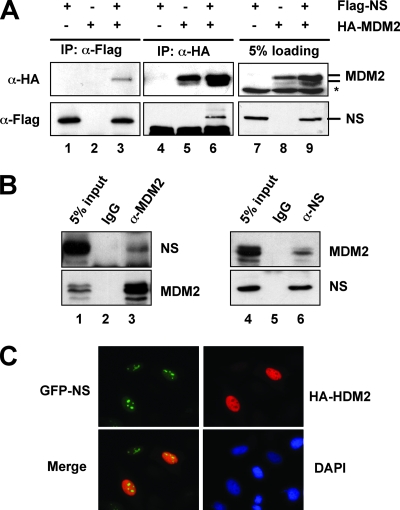
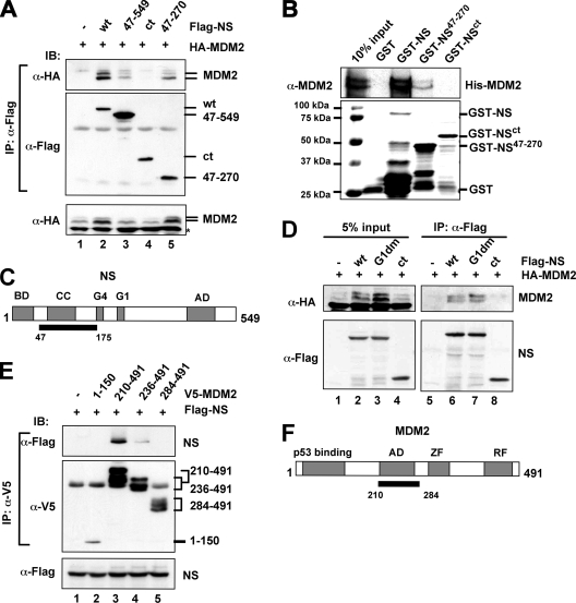
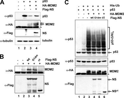
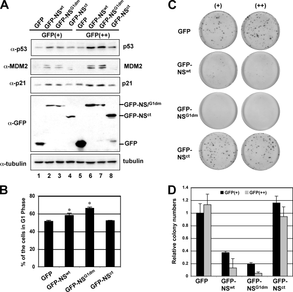
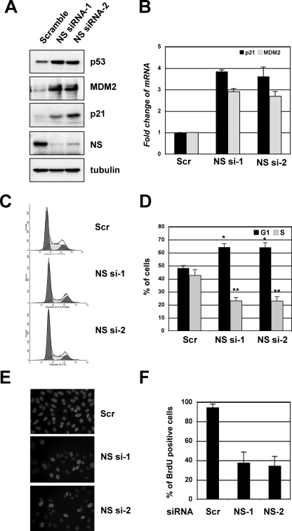
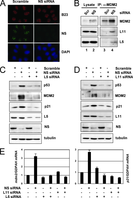
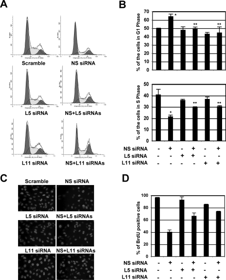
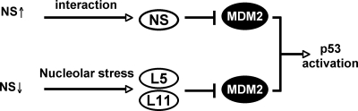
The nucleolar protein nucleostemin (NS) is essential for cell proliferation and early embryogenesis. Both depletion and overexpression of NS reduce cell proliferation. However, the mechanisms underlying this regulation are still unclear. Here, we show that NS regulates p53 activity through the inhibition of MDM2. NS binds to the central acidic domain of MDM2 and inhibits MDM2-mediated p53 ubiquitylation and degradation. Consequently, ectopic overexpression of NS activates p53, induces G(1) cell cycle arrest, and inhibits cell proliferation. Interestingly, the knockdown of NS by small interfering RNA also activates p53 and induces G(1) arrest. These effects require the ribosomal proteins L5 and L11, since the depletion of NS enhanced their interactions with MDM2 and the knockdown of L5 or L11 abrogated the NS depletion-induced p53 activation and cell cycle arrest. These results suggest that a p53-dependent cell cycle checkpoint monitors changes of cellular NS levels via the impediment of MDM2 function.


Figures

FIG. 1.
FIG. 1.
NS interacts with MDM2 in cells. (A) Ectopically expressed NS interacts with ectopically expressed MDM2 in cells. H1299 cells were transfected with Flag-NS and HA-MDM2 individually or together. Cell lysates were immunoprecipitated with anti-Flag (lanes 1 to 3) or anti-HA (lanes 4 to 6) antibodies, followed by IB with anti-Flag or anti-HA antibodies. Asterisk, nonspecific anti-HA antibody-reacting bands. (B) Endogenous NS interacts with endogenous MDM2 in cells. Cell lysates from SJSA cells were immunoprecipitated with monoclonal anti-MDM2 (4B11) antibodies (lane 3) or mouse immunoglobulin G (IgG; lane 2), polyclonal anti-NS antibodies (lane 6), or rabbit IgG (lane 5), followed by anti-MDM2 and anti-NS antibodies. (C) NS colocalizes with MDM2 in the nucleoplasm. H1299 cells were cotransfected with GFP-NS and HA-MDM2. Forty-eight hours posttransfection, the cells were immunostained with anti-MDM2 antibody, followed by staining with goat anti-mouse secondary antibody (red) and DAPI.
FIG. 2.
FIG. 2.
The coiled-coil domain-containing region of NS binds to the central acidic domain of MDM2. (A) MDM2 binds to the coiled-coil domain-containing region of NS in cells. H1299 cells were transfected with Flag-tagged full-length NS (wt) or its deletion mutants, together with the HA-MDM2 plasmid. Cell lysates were immunoprecipitated with the anti-Flag antibody, followed by IB using the indicated antibodies. The lysates were also loaded directly onto a sodium dodecyl sulfate (SDS) polyacrylamide gel for IB, using the anti-HA antibody (bottom panel). (B) MDM2 binds to NS in vitro. About 200 ng of purified GST alone, full-length GST-NS, or GST-NS deletion mutants immobilized on glutathione beads was incubated with 200 ng of His-MDM2 purified from bacteria. Bound MDM2 was detected by IB with anti-MDM2 antibodies. The GST-NS fusion proteins were visualized by Coomassie blue staining. (C) Schematic diagram of NS protein indicates the MDM2-binding domain (black bar). BD, basic domain; AD, acidic domain; CC, coiled-coil domain; G4 and G1, putative GTP binding motifs. (D) GTP binding activity of NS is not required for NS binding to MDM2. H1299 cells were transfected with HA-MDM2, together with Flag-NS, Flag-NSG1dm, Flag-NSct, or the control Flag vectors. Cell lysates were immunoprecipitated with the anti-Flag antibody, followed by IB using the indicated antibodies. The lysates were also loaded directly onto an SDS gel for IB using the indicated antibodies (left panels). (E) NS binds to the central acidic domain of MDM2. H1299 cells were transfected with plasmids encoding V5-tagged MDM2 fragments, together with the indicated Flag-NS plasmid. Cell lysates were immunoprecipitated with an anti-V5 antibody, followed by IB using the indicated antibodies. The lysates were also loaded directly onto an SDS gel for IB using anti-Flag antibody (bottom panel). (F) Schematic diagram of MDM2 protein indicating the NS binding acidic domain (AD) (black bar). ZF, zinc finger domain; RF, ring finger domain.
FIG. 3.
FIG. 3.
NS inhibits MDM2-mediated p53 ubiquitylation and degradation. (A) NS inhibits MDM2-mediated p53 degradation. H1299 cells were transfected with the indicated plasmids. Cell lysates were subjected to IB using antibodies as indicated on the left. (B) NS stabilizes MDM2 independently of p53. H1299 cells were transfected with HA-MDM2 in the presence of Flag-NS, Flag-NSG1dm, Flag-NSct, or the control Flag plasmids. Cell lysates were immunoprecipitated with the anti-Flag antibody, followed by IB using the indicated antibodies. Cell lysates were subjected to IB using the antibodies indicated on the left. (C) NS inhibits MDM2-mediated p53 ubiquitylation. H1299 cells were transfected with the indicated plasmids. The transfected cells were treated with MG132 (20 μM) for 6 h before they were harvested. Ubiquitylated p53 species were detected by IB with the anti-p53 (DO-1) antibody (upper panel). Ubiquitylated p53 [p53-(Ub)n] is indicated. The expression of total p53, MDM2, and NS proteins is shown in the lower panels. Asterisk indicates nonspecific anti-HA antibody-reacting bands.
FIG. 4.
FIG. 4.
Overexpression of NS activates p53, induces G1 arrest, and inhibits cell proliferation. (A) Overexpression of NS induces and activates p53. U2OS cells were transfected with the GFP, GFP-NS, GFP-NSG1dm, or GFP-NSct plasmid. The GFP-expressing cells were then sorted by FACS into GFP dim-positive (GFP+) and GFP bright-positive (GFP++) populations. Gating scales are shown in Fig. S2B in the supplemental material. The cell lysates were assayed by IB for the expression of p53, MDM2, p21, and GFP-NS. (B) Overexpression of NS induces G1 arrest. U2OS cells were transfected with GFP, GFP-NS, GFP-NSG1dm, or GFP-NSct. Forty-eight hours posttransfection, the GFP-expressing cells were gated for cell cycle analysis. The mean percentage of cells in G1 phase is shown. The histograms of PI staining from one representative experiment are shown in Fig. S2C in the supplemental material. Asterisk, P < 0.01, compared to that of GFP. (C and D). Overexpression of NS inhibits cell proliferation, as determined by colony formation assay. U2OS cells were transfected with the GFP, GFP-NS, GFP-NSG1dm, or GFP-NSct plasmid and sorted into GFP+ and GFP++ populations as described for panel A. Equal numbers of cells were plated and grown in media containing G418 for 3 to 4 weeks. The average numbers of colonies are shown in panel D.
FIG. 5.
FIG. 5.
Knockdown of endogenous NS activates p53 and induces G1 cell cycle arrest. (A) The knockdown of NS induces p53 levels. U2OS cells were transfected with scrambled siRNA or with one of the two NS siRNAs against different sequences. Cell lysates were assayed for the expression of p53, MDM2, p21, and NS, as indicated, by using IB assays. (B) The knockdown of endogenous NS increases the mRNA expression of the p53 target genes mdm2 and p21. Total RNAs were extracted from U2OS cells transfected with siRNAs as in panel A and subjected to RT reactions, followed by real-time PCR assays. The relative expression of the p21 and mdm2 genes was normalized against the expression of GAPDH. Scr, scrambled RNA-transfected cells. (C and D) The knockdown of endogenous NS induces G1 arrest. U2OS cells were transfected with siRNA, as described in panel A. Seventy-two hours posttransfection, the cells were harvested and stained with PI for cell cycle analysis. The histograms of PI staining from one representative experiment are shown in panel C. The mean percentages of cells in G1 or S phase are shown in panel D. *, P < 0.05; **, P < 0.01, compared to the scrambled RNA-transfected cells. (E and F) The knockdown of endogenous NS reduces cell proliferation. U2OS cells were transfected with scrambled siRNA or with one of the two above-described NS siRNAs. The cells were incubated with BrdU at 48 h posttransfection for another 20 h. The cells were then fixed and stained with anti-BrdU antibodies (red) and DAPI (blue) (E). The average of BrdU-positive cells is shown in panel F.
FIG. 6.
FIG. 6.
The knockdown of NS does not lead to nucleolar disruption, but its activation of p53 requires the ribosomal proteins L5 and L11. (A) The knockdown of endogenous NS does not disrupt the nucleolus. U2OS cells were transfected with scrambled (Scr) or NS siRNA as indicated. The cells were then stained with anti-B23 (red) and anti-NS (green) antibodies, as well as with DAPI for DNA. (B) The knockdown of endogenous NS enhances the interaction of MDM2 with L5 and L11. U2OS cells were transfected with scrambled or NS siRNA. Cell lysates were subjected to co-IP using anti-MDM2 (4B11) antibodies, followed by IB to detect the level of L11, L5, and MDM2. (C) The knockdown of L5 abolished the induction of p53 by the knockdown of NS. U2OS cells were transfected with scrambled siRNA, with NS siRNA, or with L5 siRNA as indicated. Cell lysates were subjected to IB to detect the expression of p53, MDM2, p21, L5, or NS, as indicated. (D) The knockdown of L11 abolished the induction of p53 by the knockdown of NS. U2OS cells were transfected with scrambled siRNA, with NS siRNA, or with L11 siRNA as indicated. The cell lysates were subjected to IB to detect the expression of p53, MDM2, p21, L11, or NS, as indicated. (E) The knockdown of L5 or L11 abolished the induced expression of the p21 and mdm2 mRNA by the knockdown of NS. U2OS cells were transfected with scrambled siRNA, NS siRNA, L5 siRNA, or L11 siRNA as indicated. Total RNAs were extracted and subjected to RT reactions, followed by real-time PCR assays. Relative expression of the p21 and mdm2 genes was normalized against the expression of GAPDH.
FIG. 7.
FIG. 7.
NS knockdown-induced G1 arrest requires the ribosomal proteins L5 and L11. (A and B) U2OS cells were transfected with scrambled, NS, L5, or L11 siRNA as indicated. Seventy-two hours posttransfection, the transfected cells were harvested and stained with PI for cell cycle analysis. The histograms of PI staining from one representative experiment are shown in panel A. The mean percentages of cells in G1 or S phase are shown in panel B. *, P < 0.01 compared to scrambled RNA-transfected cells; **, P < 0.01 compared to NS siRNA-transfected cells. (C and D) Knocking down L5 or L11 partially abolished the NS knockdown-mediated inhibition of cell proliferation. U2OS cells were transfected with scrambled, NS, L5, or L11 siRNA, as indicated. The cells were incubated with BrdU at 48 h posttransfection for another 20 h. The cells were then fixed and stained with anti-BrdU antibodies (red) and DAPI (blue) (E). The average of the BrdU-positive cells is shown in panel F.
FIG. 8.
FIG. 8.
Schematic model illustrating potential mechanisms underlying p53 activation by aberrantly high levels of NS upon overexpression or by siRNA-caused low levels of NS (see text for further discussion).

Similar articles

Cited by

References

    1. Ashcroft, M., Y. Taya, and K. H. Vousden. 2000. Stress signals utilize multiple pathways to stabilize p53. Mol. Cell. Biol. 203224-3233. - PMC - PubMed
    1. Banin, S., L. Moyal, S. Shieh, Y. Taya, C. W. Anderson, L. Chessa, N. I. Smorodinsky, C. Prives, Y. Reiss, Y. Shiloh, and Y. Ziv. 1998. Enhanced phosphorylation of p53 by ATM in response to DNA damage. Science 2811674-1677. - PubMed
    1. Beekman, C., M. Nichane, S. De Clercq, M. Maetens, T. Floss, W. Wurst, E. Bellefroid, and J. C. Marine. 2006. Evolutionarily conserved role of nucleostemin: controlling proliferation of stem/progenitor cells during early vertebrate development. Mol. Cell. Biol. 269291-9301. - PMC - PubMed
    1. Bhat, K. P., K. Itahana, A. Jin, and Y. Zhang. 2004. Essential role of ribosomal protein L11 in mediating growth inhibition-induced p53 activation. EMBO J. 232402-2412. - PMC - PubMed
    1. Canman, C. E., D. S. Lim, K. A. Cimprich, Y. Taya, K. Tamai, K. Sakaguchi, E. Appella, M. B. Kastan, and J. D. Siliciano. 1998. Activation of the ATM kinase by ionizing radiation and phosphorylation of p53. Science 2811677-1679. - PubMed

Publication types

MeSH terms