. 2008 Jun;28(12):3943-51.
doi: 10.1128/MCB.00013-08. Epub 2008 Apr 21.

Caspase-2 cleavage of BID is a critical apoptotic signal downstream of endoplasmic reticulum stress

Affiliations

Caspase-2 cleavage of BID is a critical apoptotic signal downstream of endoplasmic reticulum stress

John-Paul Upton et al. Mol Cell Biol. 2008 Jun.

Abstract

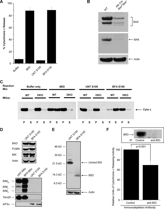
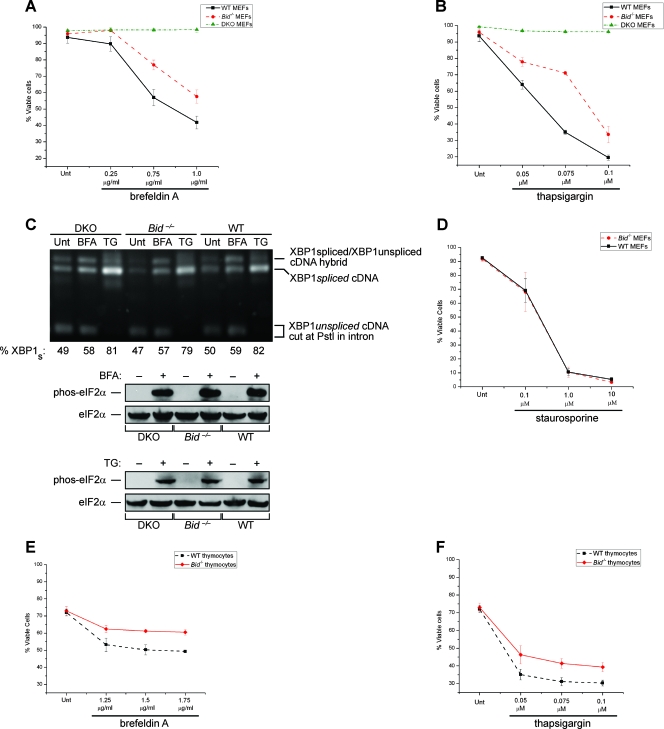
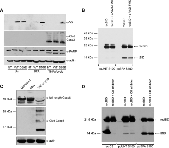
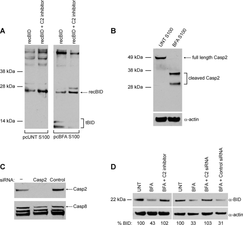
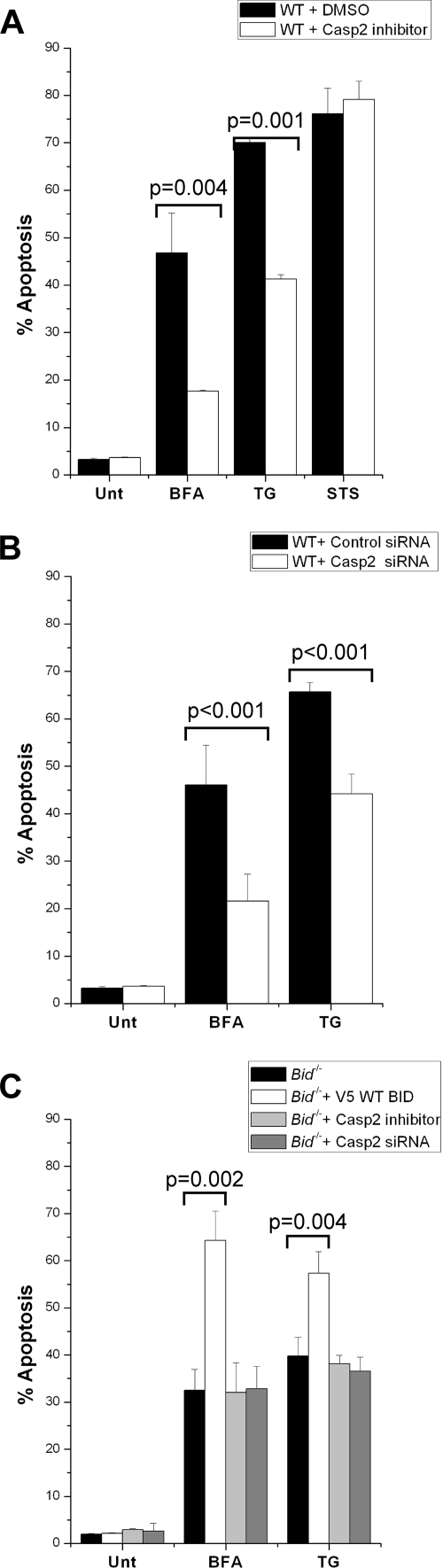
The accumulation of misfolded proteins stresses the endoplasmic reticulum (ER) and triggers cell death through activation of the multidomain proapoptotic BCL-2 proteins BAX and BAK at the outer mitochondrial membrane. The signaling events that connect ER stress with the mitochondrial apoptotic machinery remain unclear, despite evidence that deregulation of this pathway contributes to cell loss in many human degenerative diseases. In order to "trap" and identify the apoptotic signals upstream of mitochondrial permeabilization, we challenged Bax-/- Bak-/- mouse embryonic fibroblasts with pharmacological inducers of ER stress. We found that ER stress induces proteolytic activation of the BH3-only protein BID as a critical apoptotic switch. Moreover, we identified caspase-2 as the premitochondrial protease that cleaves BID in response to ER stress and showed that resistance to ER stress-induced apoptosis can be conferred by inhibiting caspase-2 activity. Our work defines a novel signaling pathway that couples the ER and mitochondria and establishes a principal apoptotic effector downstream of ER stress.


Figures

FIG. 1.
FIG. 1.
BID is proteolytically activated following ER stress and induces BAX/BAK-dependent mitochondrial cytochrome c release. (A) Cytosolic (S100) extracts from untreated (UNT) and BFA-treated DKO MEFs, as well as recombinant tBID (positive control) and reaction buffer (negative control), were incubated with isolated Jurkat mitochondria and assayed for the ability to release human cytochrome c. (B) Liver lysates (60 μg) from WT and Alb Cre Baxf/f Bak/ mice were immunoblotted for BAX and BAK. (C) S100 extracts from untreated DKO MEFs and DKO MEFs treated with 2.5 μg/ml of BFA, as well as recombinant tBID and reaction buffer, were incubated with mitochondria (Mitos) isolated from WT and DKO (Alb Cre Baxf/f Bak/) hepatocytes. The mitochondrial pellet (P) and supernatant (S) from each reaction were immunoblotted for murine cytochrome c (Cyto c). BFA-treated S100 extract releases cytochrome c from WT but not DKO mitochondria. (D) S100 extracts from untreated DKO MEFs and DKO MEFs treated with 2.5 μg/ml BFA were immunoblotted for BAD, PUMA, BIK, and actin. Mitochondrial (Mito) and S100 fractions from untreated DKO MEFs and DKO MEFs treated with 2.5 μg/ml BFA were immunoblotted for BIM, Tom20 (a mitochondrial marker), and eIF2α (a cytosolic marker). Note that no BIM was detected in the S100 fractions. BIMEL, BIM extra long; BIML, BIM long; BIMS, BIM short. (E) S100 extracts from untreated DKO MEFs and DKO MEFs treated with 2.5 μg/ml BFA were immunoblotted for BID and actin. Only tBID was detected in the BFA S100 extract. Unclvd, uncleaved. (F) S100 extract from DKO MEFs treated with 2.5 μg/ml BFA was immunodepleted with either an isotype-matched control or an anti-BID antibody and assayed for its ability to induce cytochrome c release from isolated Jurkat mitochondria. The inset shows immunodepleted extracts blotted for tBID. The relative levels of cytochrome c-releasing activity per microgram of extract were significantly reduced when BID was immunodepleted. IP, immunoprecipitation.
FIG. 2.
FIG. 2.
Bid/ cells are resistant to treatment with ER stress agents. Wild-type, Bax/ Bak/ (DKO), and Bid/ simian virus 40-transformed MEFs were treated with the indicated concentrations of either brefeldin A (A) or TG (B) for 24 and 18 h, respectively. Following these treatments, the percentages of viable (annexin V-negative) cells were determined via FACS. (C) Whole-cell lysates from DKO, Bid/, and WT MEFs that were left untreated (Unt) or treated with either 0.75 μg/ml BFA (12 h) or 0.075 μM TG (8 h) were immunoblotted for phospho (phos)-eIF2α and total eIF2α. The total RNA from DKO, Bid/, and WT MEFs that were untreated or treated with either 0.75 μg/ml BFA (4 h) or 0.075 μM TG (8 h) was isolated and subjected to an XBP1-splicing assay. The XBP1 cDNA products of PstI digestion were revealed on a 2% agarose gel. Unspliced XBP1 mRNA produced the lower doublet (291 bp and 310 bp), whereas spliced XBP1 (XBP1s) mRNA produced one 575-bp band. The highest band shown is a previously described hybrid of spliced and unspliced XBP1 cDNA fragments. Densitometry was used to determine the percent XBP1s [XBP1s/(XBP1s + XBP1us)]. (D) Bid/ and WT MEFs were treated with the indicated concentrations of STS for 18 h, and the percentages of viable (annexin V-negative) cells were determined via FACS. (E and F) Primary thymocytes from 6-week-old WT and Bid/ mice were treated with the indicated concentrations of either brefeldin A (E) or TG (F) for 8 h. Following these treatments, the percentages of viable (annexin V-negative) cells were determined via FACS. The results of all the assays were analyzed via Student's t test (n = 3). Together these data demonstrate that cells deficient in BID are resistant to apoptosis induced by ER stress agents.
FIG. 3.
FIG. 3.
ER stress induces BID cleavage at position D59 independently of caspase-8 activation. (A) Nontransfected (NT) DKO MEFS or DKO MEFS transfected with either BID-V5 (WT) or D59E BID-V5 (D59E) were left untreated or treated with 2.5 μg/ml of BFA or 1 ng/ml of TNF-α-2.5 μg/ml of cycloheximide (TNFα/cyclo) for 24 h. Whole-cell lysates immunoblotted for anti-V5 (α-V5) show that full-length WT, but not D59E BID, was reduced upon BFA treatment. Immunoblots of PARP and cleaved (Clvd) caspase-3 demonstrate that caspase-3 activity is present in TNF-α-cycloheximide-treated cells but not in BFA-treated samples. (B) S100 extracts from untreated (UNT) and BFA-treated DKO MEFs were precleared (pc) of endogenous BID/tBID and then incubated with recombinant (rec) BID with or without being pretreated with the broad-range caspase inhibitor z-VAD-fmk (40 μM). Samples were then immunoblotted for BID. Note that the S100 extract from BFA-treated DKO MEFs cleaves recBID and that this activity is not substantially reduced by z-VAD-fmk. (C) DKO MEFs were left untreated or were treated with 2.5 μg/ml BFA or 1 ng/ml of TNF-α-2.5 μg/ml of cycloheximide for 24 h. Whole-cell lysates were then immunoblotted for full-length caspase-8, cleaved caspase-8, and actin. Note that caspase-8 is cleaved and activated in response to TNF-α-cycloheximide but not to BFA. (D) S100 extracts from untreated and BFA-treated DKO MEFs were first precleared of endogenous BID/tBID and then incubated in the presence of recBID with or without pretreatment with the caspase-8 inhibitor z-IETD-fmk (40 μM). As a control, recombinant caspase-8 was incubated with recBID in the absence or presence of z-IETD-fmk (40 μM). The untreated S100 extract did not induce cleavage of recBID. The BFA extract cleaved recBID even in the presence of caspase-8 inhibition with z-IETD-fmk. α, anti.
FIG. 4.
FIG. 4.
Caspase-2 cleaves BID in response to ER stress. (A) S100 extracts from untreated (UNT) and BFA-treated DKO MEFs were precleared (pc) of endogenous BID/tBID and then incubated with recBID with or without pretreatment with the specific caspase-2 inhibitor z-VDVAD-fmk (50 μM). Samples were then immunoblotted for BID. Note that pretreatment with z-VDVAD-fmk blocked BID cleavage. (B) S100 extracts from untreated DKO MEFs and DKO MEFs treated with 2.5 μg/ml of BFA were immunoblotted for caspase-2 (Casp2) and actin. Note that BFA treatment led to caspase-2 cleavage. (C) Following a 24-h transfection of siRNA against caspase-2, immunoblotting of DKO whole-cell lysates showed that caspase-2 expression was substantially reduced. (D) DKO MEFs were left untreated or were pretreated with 50 μM z-VDVAD-fmk (C2 inhibitor), control siRNA, or caspase-2 siRNA (C2 siRNA), followed by treatment with 2.5 μg/ml of BFA for 24 h. Immunoblotting for BID and subsequent quantification by densitometry showed that inhibiting or knocking down caspase-2 protected against BFA-induced cleavage of endogenous full-length BID. α, anti.
FIG. 5.
FIG. 5.
Absence of caspase-2 (Casp2) activity confers resistance to ER stress-induced apoptosis. (A) WT MEFs were pretreated with dimethyl sulfoxide (DMSO) or 50 μM z-VDVAD-fmk (a caspase-2 inhibitor) and challenged with 0.25 μg/ml BFA (24 h), 0.05 μM TG (18 h), or 1.0 μM STS (18 h). Cells were then harvested and analyzed by FACS for annexin V staining. Note that cells pretreated with z- VDVAD-fmk were significantly resistant to the ER stress agents but not to STS. (B) WT MEFs were transfected with either 2 μM control siRNA or caspase-2 siRNA and treated with 0.25 μg/ml of BFA (24 h) or 0.05 μM of TG (18 h). Cells were then harvested and analyzed by FACS for annexin V staining. Note that cells transfected with caspase-2 siRNA were significantly resistant to both of these ER stress agents. (C) Bid/ MEFs were either pretreated with 50 μM z-VDVAD-fmk (6 h) or transfected with 1 μg V5-tagged WT BID or 2 μM caspase-2 siRNA and then challenged with 0.75 μg/ml of BFA (24 h) or 0.075 μM of TG (18 h). The cells were then harvested and analyzed by FACS for annexin V staining. Note that pharmacological inhibition or siRNA knockdown of caspase-2 did not further protect Bid/ cells against ER stress agents. The results of all the assays were analyzed via Student's t test (n = 3).

References

    1. Bonzon, C., L. Bouchier-Hayes, L. J. Pagliari, D. R. Green, and D. D. Newmeyer. 2006. Caspase-2-induced apoptosis requires Bid cleavage: a physiological role for Bid in heat shock-induced death. Mol. Biol. Cell 172150-2157. - PMC - PubMed
    1. Calfon, M., H. Zeng, F. Urano, J. H. Till, S. R. Hubbard, H. P. Harding, S. G. Clark, and D. Ron. 2002. IRE1 couples endoplasmic reticulum load to secretory capacity by processing the XBP-1 mRNA. Nature 41592-96. - PubMed
    1. Cheung, H. H., N. Lynn Kelly, P. Liston, and R. G. Korneluk. 2006. Involvement of caspase-2 and caspase-9 in endoplasmic reticulum stress-induced apoptosis: a role for the IAPs. Exp. Cell Res. 3122347-2357. - PubMed
    1. Chipuk, J. E., L. Bouchier-Hayes, and D. R. Green. 2006. Mitochondrial outer membrane permeabilization during apoptosis: the innocent bystander scenario. Cell Death Differ. 131396-1402. - PubMed
    1. Credle, J. J., J. S. Finer-Moore, F. R. Papa, R. M. Stroud, and P. Walter. 2005. On the mechanism of sensing unfolded protein in the endoplasmic reticulum. Proc. Natl. Acad. Sci. USA 10218773-18784. - PMC - PubMed

Publication types

MeSH terms

Substances