Review
. 2008 May;18(5):538-48.
doi: 10.1038/cr.2008.52.

Genomic analysis of epithelial ovarian cancer

Affiliations
Review

Genomic analysis of epithelial ovarian cancer

John Farley et al. Cell Res. 2008 May.

Abstract

Ovarian cancer is a major health problem for women in the United States. Despite evidence of considerable heterogeneity, most cases of ovarian cancer are treated in a similar fashion. The molecular basis for the clinicopathologic characteristics of these tumors remains poorly defined. Whole genome expression profiling is a genomic tool, which can identify dysregulated genes and uncover unique sub-classes of tumors. The application of this technology to ovarian cancer has provided a solid molecular basis for differences in histology and grade of ovarian tumors. Differentially expressed genes identified pathways implicated in cell proliferation, invasion, motility, chromosomal instability, and gene silencing and provided new insights into the origin and potential treatment of these cancers. The added knowledge provided by global gene expression profiling should allow for a more rational treatment of ovarian cancers. These techniques are leading to a paradigm shift from empirical treatment to an individually tailored approach. This review summarizes the new genomic data on epithelial ovarian cancers of different histology and grade and the impact it will have on our understanding and treatment of this disease.


Figures

Figure 1
Figure 1
Photomicrograph of papillary serous ovarian cancer: low malignant potential tumors (A: low power, B: high power) and high-grade invasive carcinoma (C: low power, D: high power).
Figure 2
Figure 2
Model of the development of an epithelial ovarian tumor. Incessant ovulation and wound repair increases the risk of genetic abnormalities, leading to dysplastic changes in epithelial cells lining the mullerian inclusion cyst. Stromal microenvironment in the forms of activated fibroblast formation, microvessel proliferation, and growth factors contributes to dysplastic formation and eventual malignant transformation.
Figure 3
Figure 3
Principle component analysis (PCA) of the expression profiles of ovarian specimens. Samples with similar profiles cluster relatively close. (A) PCA of the normal groups. (B) Unsupervised MDS of the normal groups and the serous ovarian carcinoma samples.
Figure 4
Figure 4
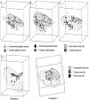
Principle component analysis (PCA) of ovarian and endometrial cancers according to histology. (A) PCA of tumors with serous histology showing two nonoverlapping elliptical regions separating endometrial (top) from ovarian (bottom) specimens. (B) PCA of tumors with endometrioid histology showing two nonoverlapping elliptical regions separating endometrial (top) from ovarian (bottom) specimens. (C) PCA of tumors with clear cell histology showing overlapping elliptical regions representing endometrial (top) and ovarian (bottom) specimens. (D) PCA of tumors according to organ of origin shows three overlapping elliptical regions among ovarian, endometrial, and renal clear cell specimens, with two different orientations (1 and 2).
Figure 5
Figure 5
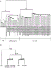
(A) Unsupervised hierarchical clustering analysis of LMP, low-grade, high-grade, and OSE specimens. OSE, LMP, and low-grade tumors grouped along node A and early-stage and late-stage high-grade lesions grouped along node B. OSE clustered separately from LMP and low-grade tumors. LMP and low-grade tumors were indistinguishable from one another. Low-grade and early-stage high-grade samples are indicated in bold. Misclassified specimens are bold italicized. (B) Binary tree prediction followed by leave-one-out cross-validation to estimate the error associated with the tree-building process. LMP tumors and low-grade cancers were more closely aligned to each other, indicated by the high misclassification rate (30.8%), but are distinct from the early-stage and late-stage high-grade tumors with a low misclassification rate (3.7%). Percentages indicate the misclassification error associated with each node.

References

    1. Jemal A, Siegel R, Ward E, et al. Cancer statistics, 2008. CA Cancer J Clin 2008; 58:71–96. - PubMed
    1. Cannistra SA. Cancer of the ovary. N Engl J Med 2004; 351:2519–2529. - PubMed
    1. Bhoola S, Hoskins WJ. Diagnosis and management of epithelial ovarian cancer. Obstet Gynecol 2006; 107:1399–1410. - PubMed
    1. Ozols RF, Rubin SC, Thomas GM, Robboy SJ. Epithelial ovarian cancer In: Hoskins WJ, Young RC, Markman M, Perez CA, Barakat R, Randall M, eds. Principles and Practice of Gynecologic Oncology. Philadelphia, PA: Lippincott Williams & Wilkins, 2005:895–988.
    1. Yawn BP, Barrette BA, Wollan PC. Ovarian cancer: the neglected diagnosis. Mayo Clin Proc 2004; 79:1277–1282. - PubMed