. 2007:105:121-9; discusion 129-31.

Macular holes and macular pucker: the role of vitreoschisis as imaged by optical coherence tomography/scanning laser ophthalmoscopy

Affiliations

Macular holes and macular pucker: the role of vitreoschisis as imaged by optical coherence tomography/scanning laser ophthalmoscopy

Jerry Sebag et al. Trans Am Ophthalmol Soc. 2007.

Abstract

Purpose: The pathogenesis of macular pucker and macular holes is poorly understood. Anomalous posterior vitreous detachment (PVD) and vitreoschisis have been proposed as possible mechanisms. This study used clinical imaging to seek vitreoschisis and study the topographic features of macular pucker and macular holes.

Methods: Combined optical coherence tomography and scanning laser ophthalmoscopy (OCT/SLO) was performed in 45 eyes with macular hole and 44 eyes with macular pucker. Longitudinal imaging was used to identify vitreoschisis and measure retinal thickness. The topographic features of eyes with macular hole with eccentric macular contraction were compared to 24 eyes with unifocal macular pucker using coronal plane imaging.

Results: Vitreoschisis was detected in 24 of 45 eyes (53.3%) with macular hole and 19 of 44 (43.2%) with macular pucker. Retinal contraction was detected eccentrically in the macula of 18 of 45 eyes (40%) with macular hole. In eyes with macular hole with unifocal retinal contraction, the average surface area of contraction (23.12 +/- 18.79 mm(2)) was significantly smaller than in eyes with macular pucker (63.20 +/- 23.68 mm(2); P = .006). The distance from the center of retinal contraction to the center of the macula was significantly greater in eyes with macular hole (8.64 +/- 2.33 mm) than eyes with macular pucker (4.45 +/- 1.90 mm; P = .0001).

Conclusion: Vitreoschisis was detected in about half of all eyes with macular hole and macular pucker. The topographic and structural features in eyes with macular hole with retinal contraction differed in comparison to eyes with macular pucker alone, suggesting that although each condition may begin with anomalous PVD, differences in subsequent cell migration and proliferation probably result in the different clinical appearances detected in this study.


Figures

FIGURE 1
FIGURE 1
Analysis of topographic features in macular pucker. A, Grayscale SLO image of the fundus in a subject’s left eye. B, Overlay of the coronal OCT image (color photo C) superimposed on the SLO fundus image (grayscale photo A) with point-to-point registration. C, Coronal plane OCT image, in color. The overlay image (B) was used to characterize the macular hole and/or macular pucker (as in this case) for topographic analysis.
FIGURE 2
FIGURE 2
Intersecting planes analysis of macular hole. Three-dimensional reconstruction of a transverse OCT image at 45 degrees intersecting with the SLO fundus image of a patient with a macular hole. The point that was identified as the center of contraction on the fundus/SLO overlay corresponded to the same point on OCT, which was used to perform retinal thickness measurement.
FIGURE 3
FIGURE 3
Quantitative analysis of retinal contraction in macular hole. Longitudinal OCT imaging was performed at an axis of 45 degrees (red line with arrowhead on the small grayscale SLO images of fundus; to the right). A, White arrow points to the center of eccentric retinal contraction identified by 3-dimensional rendering. B, White arrow demonstrates the vertical caliper function used to measure retinal thickness (272 μm at that point).
FIGURE 4
FIGURE 4
Quantitative analysis of coronal plane OCT images in macular holes. Measurement of the distance from the center of contraction to the center of the macular hole and the diameter of retinal contraction area was performed using Adobe Photoshop software. The white line is a representation of the ruler function used to measure the distance between the macular hole and the center of the macular pucker. The blue line is a representation of the ruler function used to measure the diameter of the contraction center for calculation of contraction area.
FIGURE 5
FIGURE 5
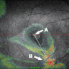
Retinal contraction in macular holes. Coronal plane image (color) superimposed on grayscale SLO fundus image of a macular hole (A) with an eccentric focus of macular pucker (B).
FIGURE 6
FIGURE 6
Vitreoschisis in macular hole. Longitudinal OCT and SLO imaging of the vitreoretinal interface in an eye with a stage 3 macular hole demonstrates the two walls (inner wall is anterior, toward the top of the photo; outer wall is posterior, toward the bottom of the photo, attached to the inner surface of the retina) of vitreoschisis.
FIGURE 7
FIGURE 7
Vitreoschisis in macular pucker. Longitudinal OCT and SLO (right) imaging of the vitreoretinal interface demonstrates the two walls (inner wall is anterior, toward the top of the photo; outer wall is posterior, toward the bottom of the photo) of vitreoschisis forming a “lambda” sign where they rejoin.
FIGURE 8
FIGURE 8
Lamellar structure of posterior vitreous cortex. Immufluorescent micrograph of human vitreoretinal interface. Using anti-ABA lectin binding, the internal limiting lamina stains intensely as a horizontal line in the middle of this photograph, and the lamellae of the posterior vitreous cortex are apparent anterior to this line. (Courtesy of Dr Greg Hageman, University of Iowa)
FIGURE 9
FIGURE 9
Top left, Human hyalocytes in situ. Dark-field slit microscopy of the posterior vitreous in a 59-year-old man. Arrow indicates hyalocytes embedded within the posterior vitreous cortex. Top right, Explanted human posterior vitreous cortex with hyalocytes. Phase contrast microscopy of hyalocytes in the posterior vitreous cortex of an 11-year-old girl. The specimen was fresh and unfixed (original magnification, ×290). (Specimen courtesy of New England Eye Bank). Bottom, Ultrastructure of human hyalocyte. Transmission electron micrcograph of human hyalocyte embedded in the dense collagen matrix of the posterior vitreous cortex (black C) (original magnification, ×11,670). Mi, microvilli, typical of phagocytes; N, lobulated nucleus with dense marginal chromatin (white C); M, mitochondria; V, vacuole; black arrows indicate cytoplasmic dense granules. (Courtesy of Joe Craft and Dr Daniel Albert, Cogan Laboratory of Ophthalmic Pathology, Harvard Medical School)

References

    1. Sebag J. Anomalous posterior vitreous detachment: a unifying concept in vitreoretinal disease. Graefes Arch Clin Exp Ophthalmol. 2004;242:690–698. - PubMed
    1. Messmer EM, Heidenkummer H, Kampic A. Ultrastructure of epiretinal membranes associated with macular holes. Graefes Arch Clin Exp Ophthalmol. 1998;236:248–254. - PubMed
    1. Chen TY, Yang CM, Liu KR. Intravitreal triamcinalone staining observation of residual undetached cortical vitreous after posterior vitreous detachment. Eye. 2006;20:423–427. - PubMed
    1. Snead DRJ, Cullen S, James S, et al. Hyperconvolution of the inner limiting membrane in vitreomaculopathies. Graefes Arch Clin Exp Ophthalmol. 1998;236:248–254. - PubMed
    1. Noyes HD. Detachment of the retina, with laceration at the macula lutea. Trans Am Ophthalmol Soc. 1871;1:128. - PMC - PubMed