. 2007:105:344-64.

A reconsideration of anterior chamber flare and its clinical relevance for children with chronic anterior uveitis (an American Ophthalmological Society thesis)

Affiliations

A reconsideration of anterior chamber flare and its clinical relevance for children with chronic anterior uveitis (an American Ophthalmological Society thesis)

Gary N Holland. Trans Am Ophthalmol Soc. 2007.

Abstract

Purpose: To gain a better understanding of laser flare photometry values ("flare") as a feature of chronic anterior uveitis in children; to identify relationships between flare and other patient and disease characteristics; to describe changes in flare during course of disease; and specifically to determine whether elevated flare is predictive of subsequent adverse events.

Methods: A retrospective review of medical records was performed for all children (aged < or =16 years at disease onset) with chronic anterior uveitis presumed to be noninfectious, who were examined by one clinician at the Jules Stein Eye Institute since laser flare photometry became available at that facility. All involved eyes were studied. Cross-sectional analysis compared initial flare to other characteristics. Relationships between potential risk factors and outcomes were studied by Kaplan-Meier analyses and Cox proportional hazards regression models.

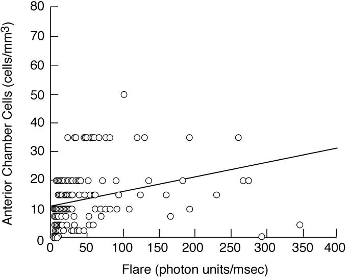
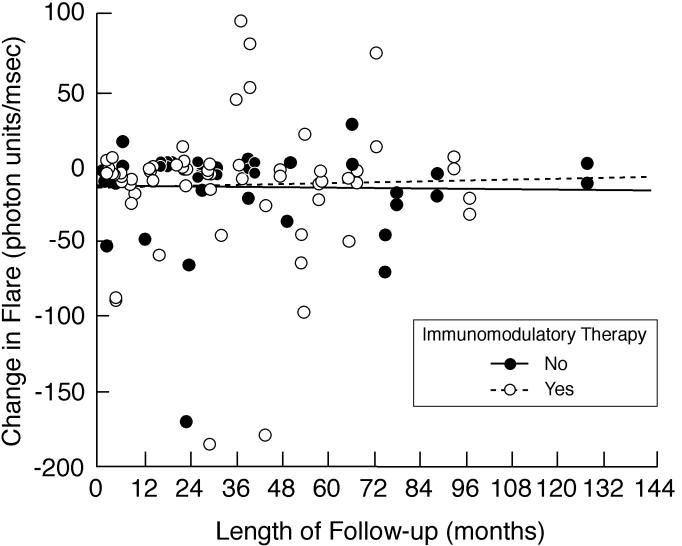
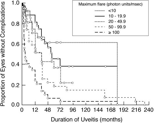
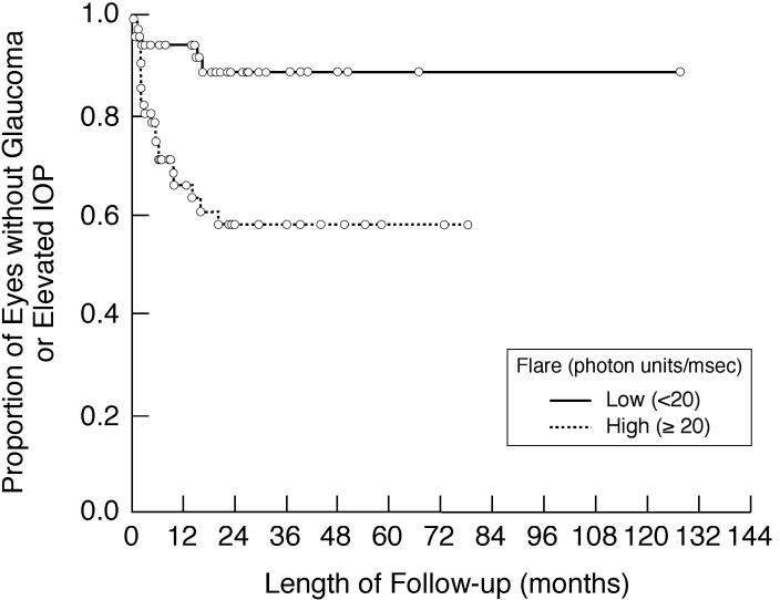
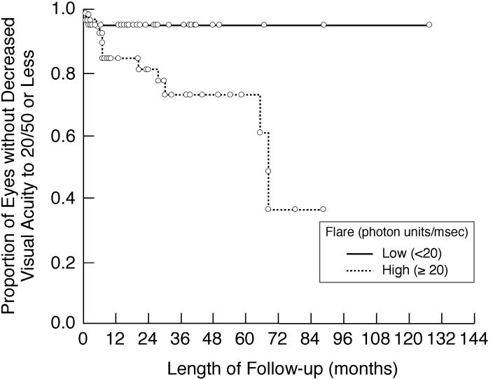
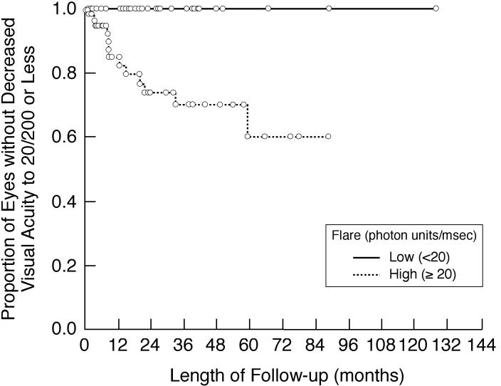
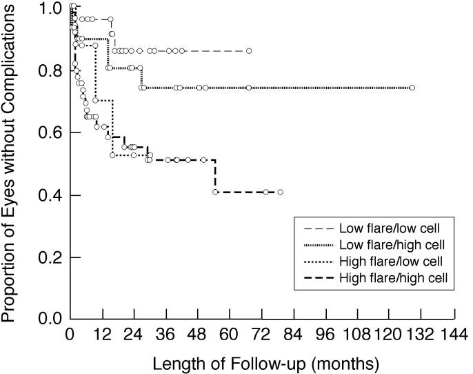
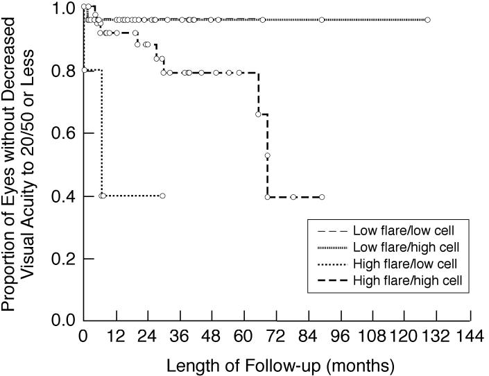
Results: Included were 114 patients (198 involved eyes). Follow-up ranged from 0 to 154.8 months (median, 23.5 months for 82 patients with follow-up). Flare was related to the following factors: anterior chamber cells, keratic precipitates, papillitis, and various complications of uveitis, including band keratopathy, posterior synechiae, and cataract. Flare was not a function of disease duration. High flare was associated with an increased risk of vision loss and development of new vision-threatening complications, including glaucoma/increased intraocular pressure, during follow-up; risk was independent of anterior chamber cells.

Conclusions: Flare may be useful in the evaluation and management of chronic anterior uveitis in children. Flare is a marker of disease severity and is predictive of adverse events during the course of disease.


Figures

FIGURE 1
FIGURE 1
Scatter plot showing baseline/first flare value vs number of anterior chamber cells from the same examination date for eyes of children with chronic anterior uveitis. See text for explanation of conversion from categorical cell score to numerical cell value.
FIGURE 2
FIGURE 2
Scatter plot showing baseline/first flare value vs most recent flare value for eyes of children with chronic anterior uveitis. There was little overall change in flare values during the course of disease, regardless of length of follow-up.
FIGURE 3
FIGURE 3
Scatter plot showing length of follow-up vs change in flare values for eyes of children with chronic anterior uveitis. The difference was determined by subtracting baseline or first flare value from the most recent flare value. There was an overall decline in flare during the course of disease for involved eyes for children with follow-up. Shown are subgroup analyses based on use of immunomodulatory therapy.
FIGURE 4
FIGURE 4
Kaplan-Meier analysis showing duration of uveitis vs the proportion of involved eyes of children with chronic anterior uveitis that are without any vision-threatening complications. Shown are subgroup analyses based on maximum flare value (<10 pu/msec; 10–19.9 pu/msec; 20–49.9 pu/msec; 50–99.9 pu/msec; ≥100 pu/msec). For study purposes, complications identified at baseline were assumed to have occurred at baseline. Duration of uveitis for each patient is from onset of disease, as determined by history.
FIGURE 5
FIGURE 5
Kaplan-Meier analysis showing the proportion of involved eyes of children with chronic anterior uveitis that are without any vision-threatening complication during follow-up after initial flare determination. Shown are subgroup analyses based on baseline/first flare value (<20 pu/msec vs 20 ≥pu/msec).
FIGURE 6
FIGURE 6
Kaplan-Meier analysis showing the proportion of involved eyes of children with chronic anterior uveitis that are without new evidence of glaucoma or loss of intraocular pressure control during follow-up after initial flare determination. Shown are subgroup analyses based on baseline/first flare value (<20 pu/msec vs 20 ≥pu/msec). IOP, intraocular pressure.
FIGURE 7
FIGURE 7
Kaplan-Meier analysis showing the proportion of involved eyes of children with chronic anterior uveitis that are without decreased visual acuity to 20/50 or less during follow-up after initial flare determination. Shown are subgroup analyses based on baseline/first flare value (<20 pu/msec vs 20 ≥pu/msec).
FIGURE 8
FIGURE 8
Kaplan-Meier analysis showing the proportion of involved eyes of children with chronic anterior uveitis that are without decreased visual acuity to 20/200 or less during follow-up after initial flare determination. Shown are subgroup analyses based on baseline/first flare value (<20 pu/msec vs 20 ≥pu/msec).
FIGURE 9
FIGURE 9
Kaplan-Meier analysis showing the proportion of involved eyes of children with chronic anterior uveitis that are without any vision-threatening complications during follow-up after initial flare determination. Shown are subgroup analyses based on baseline/first flare and corresponding anterior chamber cell score. Low flare is defined as <20 pu/msec; high flare is defined as ≥20 pu/msec. Low cells is defined as <1+; high cells is defined as ≥1+.
FIGURE 10
FIGURE 10
Kaplan-Meier analysis showing the proportion of involved eyes of children with chronic anterior uveitis that are without decreased visual acuity to 20/50 or less during follow-up after initial flare determination. Shown are subgroup analyses based on baseline/first flare and corresponding anterior chamber cell score. Low flare is defined as <20 pu/msec; high flare is defined as ≥20 pu/msec. Low cells is defined as <1+; high cells is defined as ≥1+.

References

    1. Hogan MJ, Kimura SJ, Thygeson P. Signs and symptoms of uveitis. I. Anterior uveitis. Am J Ophthalmol. 1959;47:155–170. - PubMed
    1. Duke-Elder S, Perkins ES. The clinical signs and symptoms of uveitis. In: Duke-Elder S, editor. System of Ophthalmology. St Louis: CV Mosby; 1966. pp. 132–150.
    1. Schlaegel TF. Essentials of Uveitis. 1. Boston: Little, Brown; 1967. p. 7.
    1. Smith RE, Nozik RA. Uveitis. A Clinical Approach to Diagnosis and Management. 2. Balitmore: Williams & Wilkins; 1989. Signs of uveitis; pp. 16–17.
    1. Whitcup SM. Examination of the patient with uveitis. In: Nussenblatt RB, Whitcup SM, editors. Uveitis. Fundamentals and Clinical Practice. 3. Philadelphia: Mosby; 2004. pp. 57–69.

Publication types