. 2007:105:530-63.

Endothelial keratoplasty: clinical outcomes in the two years following deep lamellar endothelial keratoplasty (an American Ophthalmological Society thesis)

Endothelial keratoplasty: clinical outcomes in the two years following deep lamellar endothelial keratoplasty (an American Ophthalmological Society thesis)

Mark A Terry. Trans Am Ophthalmol Soc. 2007.

Abstract

Purpose: To evaluate the clinical outcome of small-incision, deep lamellar endothelial keratoplasty (DLEK) for the treatment of endothelial dysfunction.

Methods: A prospective series of 79 eyes that underwent DLEK by a single surgeon was evaluated. Best spectacle-corrected visual acuity (BSCVA), refractive astigmatism, and central endothelial cell density (ECD) were measured preoperatively and at 6, 12, and 24 months.

Results: Data was available on 78 eyes (99%) at 6 months, 77 eyes (97%) at 1 year, and 79 eyes (100%) at 2 years. Mean BSCVA preoperatively of 20/71 improved to 20/42 by 6 months and remained stable. Eliminating eyes with known retinal disease, BSCVA of 20/40 or better was present in 60% (40 of 67) of eyes at 6 months, 74% (49 of 66) of eyes at 1 year, and 79% (53 of 68) of eyes at 2 years. Refractive astigmatism preoperatively was .91 +/-.78 diopters and was unchanged by surgery over time with results at 6 months of 1.11 +/-.76 (P = .052, power = .43), 1 year 1.04 +/-.80 (P =.287, power = .06), and 2 years 1.10 +/-.70 (P =.467, power = .22). The mean donor ECD preoperatively was 2819 +/- 225 (2389 to 3385) cells/mm(2), and this decreased by 26% at 6 months (2095 +/- 380) (1097 to 2920) (P = .0001; 95% confidence interval [CI] = 643-809), 3% fewer at 1 year (2009 +/- 393) (612 to 2723) (P = .054, power = .5), and 17% fewer at 2 years (1536 +/- 547) (500 to 2546) (P < .001, 95% CI = 368-585). Complications included one primary graft failure and 4 dislocations into the anterior chamber.

Conclusions: DLEK provides improved vision and minimal refractive astigmatic change, but progressive ECD decrease over time is of concern.


Figures
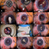
FIGURE 1
FIGURE 1
Surgical steps for small-incision deep lamellar endothelial keratoplasty (DLEK). Top row: Left, Surface of cornea is marked with an 8.0-mm circular template mark after anterior chamber is filled with Healon viscoelastic. Center, A 5.0-mm limbal scleral incision is made temporally, and a deep scleral cornea pocket is created. Right, A semisharp broad stromal dissector extends the deep lamellar pocket to the midpupillary zone. Second row: Left, A curved semisharp dissector extends the deep lamellar pocket distally to create a pocket 1 mm wider in diameter than the overlying template mark. Center, Specialized highly curved, low profile scissors are used to excise the posterior tissue, following the path of the overlying circular template mark. Right, All viscoelastic is removed from the eye with an irrigation-aspiration cannula. Third row: Left, Donor tissue is mounted onto an artificial anterior chamber, and after a 350-μm-deep limbal incision is made, the deep stromal dissection pocket is created, limbus to limbus. Center, The donor tissue is placed endothelial side up, punched with an 8.0-mm donor trephine, a Healon strip is placed on the central endothelium, and then the tissue is folded into a 60%/40% “taco” shape. Right, The donor tissue is inserted into the anterior chamber using special noncrushing forceps. Bottom row: Left, Irrigation from the paracentesis site deepens the chamber and the tissue unfolds spontaneously. Center, With the chamber filled with air and the wound closed, a reverse Sinskey hook is used to pull the recipient bed edges posterior to the donor edges. Right, At the close of the procedure, a 4-mm diameter or smaller air bubble is left in the anterior chamber, and a collagen shield with antibiotics and steroids is placed over the cornea and the eye patched until the next morning.
FIGURE 2
FIGURE 2
Deep lamellar endothelial keratoplasty (DLEK) surgery for the treatment of edema due to blunt trauma. Left, Preoperative appearance of severe edema following blunt trauma. Note extensive pigment coating posterior cornea accentuating the striate keratopathy. Preoperative vision was counting fingers at 2 feet. Right, Postoperative appearance of cornea with central clarity enabling subsequent cataract surgery. Peripheral surface pannus vessels are seen in the superior nasal quadrant. Spectacle visual acuity at 2 years after DLEK surgery (and after cataract surgery) was 20/30.
FIGURE 3
FIGURE 3
Surface scar from herpes simplex infection following deep lamellar endothelial keratoplasty (DLEK) surgery. Note the clarity of the graft central to the surface scar. Visual acuity was 20/30.
FIGURE 4
FIGURE 4
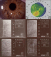
Small-incision deep lamellar endothelial keratoplasty (DLEK): Eye 35 in series. Top row: Left, Crystal clear DLEK cornea 3 years after surgery. Right, Normal corneal topography with 0.35 diopters of cylinder 3 years after surgery. Center row: Left, Endothelial cell density at 6 months postoperatively = 2329 cells/mm2. Right, Endothelial cell density at 12 months postoperatively = 2357 cells/mm2. Bottom row: Left, Endothelial cell density at 24 months postoperatively = 1938 cells/mm2. Right, Endothelial cell density at 36 months postoperatively = 864 cells/mm2.

References

    1. Zirm E. Eine erfolgreiche totale Keratoplastik. Arch Ophthalmol. 1906;64:580–593.
    1. Zirm ME, Mannis AA. Eduard Zirm 1863–1944. In: Mannis MJ, Mannis AA, editors. History of Ophthalmology—Corneal Transplantation: A History in Profiles. Vol. 6. Belgium: J.P. Wayenborgh; 1999. pp. 129–141.
    1. Sugar A, Sugar J. Techniques in penetrating keratoplasty: a quarter century of development. Cornea. 2000;19:603–610. - PubMed
    1. Melles GR, Lander F, van Dooren BR, et al. Preliminary clinical results of posterior lamellar keratoplasty through a sclerocorneal pocket incision. Ophthalmology. 2000;107:1850–1856. - PubMed
    1. Terry MA. Deep lamellar endothelial keratoplasty (DLEK): pursuing the ideal goals of endothelial replacement. EYE. 2003;17:982–988. - PubMed

Publication types