

Three-dimensional ontogenetic shape changes in the human cranium during the fetal period
- PMID: 18430090
- PMCID: PMC2409084
- DOI: 10.1111/j.1469-7580.2008.00884.x
Three-dimensional ontogenetic shape changes in the human cranium during the fetal period
Abstract
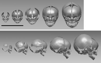
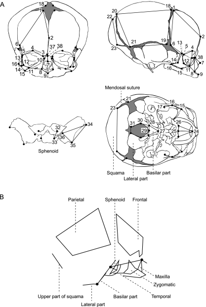
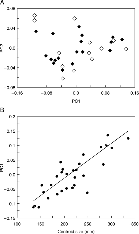
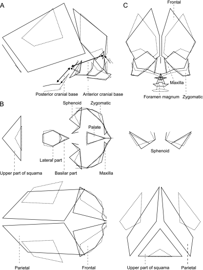
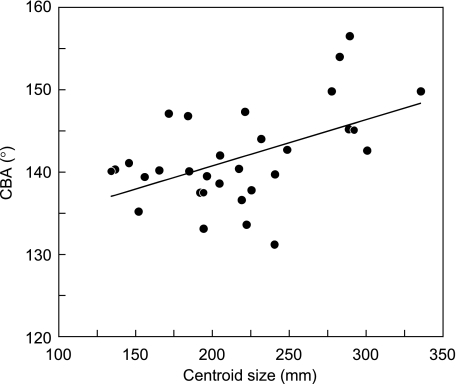
Knowledge of the pattern of human craniofacial development in the fetal period is important for understanding the mechanisms underlying the emergence of variations in human craniofacial morphology. However, the precise character of the prenatal ontogenetic development of the human cranium has yet to be fully established. This study investigates ontogenetic changes in cranial shape in the fetal period, as exhibited in Japanese fetal specimens housed at Kyoto University. A total of 31 human fetal specimens aged from approximately 8 to 42 weeks of gestation underwent helical computed tomographic scanning, and 68 landmarks were digitized on the internal and external surfaces of the extracted crania. Ontogenetic shape change was then analyzed cross-sectionally and three-dimensionally using a geometric morphometric technique. The results of the present study are generally consistent with previously reported findings. It was found that during the prenatal ontogenetic process, the growth rate of the length of the cranium is greater than that of the width and height, and the growth rate of the length of the posterior cranial base is smaller than that of the anterior cranial base. Furthermore, it was observed that the change in shape of the human viscerocranium is smaller than that of the neurocranium during the fetal period, and that concurrently the basicranium extends by approximately 8 degrees due to the relative elevation of the basilar and lateral parts of occipital bone. These specific growth-related changes are the opposite of those reported for the postnatal period. Our findings therefore indicate that the allometric pattern of the human cranium is not a simple continuous transformation, but changes drastically from before to after birth.
Figures

Similar articles
-
Prenatal ontogeny of subspecific variation in the craniofacial morphology of the Japanese macaque (Macaca fuscata).Primates. 2010 Jul;51(3):263-71. doi: 10.1007/s10329-010-0197-3. Epub 2010 Apr 2. Primates. 2010. PMID: 20361348
-
Craniofacial levels and the morphological maturation of the human skull.J Anat. 2006 Nov;209(5):637-54. doi: 10.1111/j.1469-7580.2006.00644.x. J Anat. 2006. PMID: 17062021 Free PMC article.
-
Allometries throughout the late prenatal and early postnatal human craniofacial ontogeny.Anat Rec (Hoboken). 2007 Sep;290(9):1112-20. doi: 10.1002/ar.20581. Anat Rec (Hoboken). 2007. PMID: 17721983
-
Growth-related shape changes in the fetal craniofacial complex of humans (Homo sapiens) and pigtailed macaques (Macaca nemestrina): a 3D-CT comparative analysis.Am J Phys Anthropol. 2003 Apr;120(4):339-51. doi: 10.1002/ajpa.10125. Am J Phys Anthropol. 2003. PMID: 12627529
-
Growth movements during prenatal development of human facial morphology.Prog Clin Biol Res. 1985;187:57-66. Prog Clin Biol Res. 1985. PMID: 3903766 Review.
Cited by
-
Size, shape, and form: concepts of allometry in geometric morphometrics.Dev Genes Evol. 2016 Jun;226(3):113-37. doi: 10.1007/s00427-016-0539-2. Epub 2016 Apr 1. Dev Genes Evol. 2016. PMID: 27038023 Free PMC article. Review.
-
Age-at-Death Estimation of Fetuses and Infants in Forensic Anthropology: A New "Coupling" Method to Detect Biases Due to Altered Growth Trajectories.Biology (Basel). 2022 Jan 27;11(2):200. doi: 10.3390/biology11020200. Biology (Basel). 2022. PMID: 35205067 Free PMC article.
-
Morphometric study of the primary ossification center of the frontal squama in the human fetus.Surg Radiol Anat. 2020 Jul;42(7):733-740. doi: 10.1007/s00276-020-02425-7. Epub 2020 Feb 5. Surg Radiol Anat. 2020. PMID: 32025797 Free PMC article.
-
Ultrasonographic study of fetal facial profile markers during the first trimester.BMC Pregnancy Childbirth. 2021 Apr 24;21(1):324. doi: 10.1186/s12884-021-03813-6. BMC Pregnancy Childbirth. 2021. PMID: 33894762 Free PMC article.
-
The influence of life history and sexual dimorphism on entheseal changes in modern humans and African great apes.PLoS One. 2014 Sep 24;9(9):e107963. doi: 10.1371/journal.pone.0107963. eCollection 2014. PLoS One. 2014. PMID: 25251439 Free PMC article.
References
-
- Ackermann RR, Krovitz GE. Common patterns of facial ontogeny in the hominid lineage. Anat Rec. 2002;269:142–147. - PubMed
-
- Birch RH. Foetal retrognathia and the cranial base. Angle Orthod. 1968;38:231–235. - PubMed
-
- Bookstein FL, Gunz P, Mitteroecker P, Prossinger H, Schaefer K, Seidler H. Cranial integration in Homo: singular warps analysis of the midsagittal plane in ontogeny and evolution. J Hum Evol. 2003;44:167–187. - PubMed
-
- Bulygina E, Mitteroecker P, Aiello L. Ontogeny of facial dimorphism and patterns of individual development within one human population. Am J Phys Anthropol. 2006;131:432–443. - PubMed
-
- Burdi AR. Cephalometric growth analyses of the human upper face region during the last two trimesters of gestation. Am J Anat. 1969;125:113–122. - PubMed
Publication types
MeSH terms
LinkOut - more resources
Full Text Sources
