

Systemic 5-fluorouracil treatment causes a syndrome of delayed myelin destruction in the central nervous system
- PMID: 18430259
- PMCID: PMC2397490
- DOI: 10.1186/jbiol69
Systemic 5-fluorouracil treatment causes a syndrome of delayed myelin destruction in the central nervous system
Abstract
Background: Cancer treatment with a variety of chemotherapeutic agents often is associated with delayed adverse neurological consequences. Despite their clinical importance, almost nothing is known about the basis for such effects. It is not even known whether the occurrence of delayed adverse effects requires exposure to multiple chemotherapeutic agents, the presence of both chemotherapeutic agents and the body's own response to cancer, prolonged damage to the blood-brain barrier, inflammation or other such changes. Nor are there any animal models that could enable the study of this important problem.
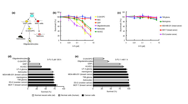
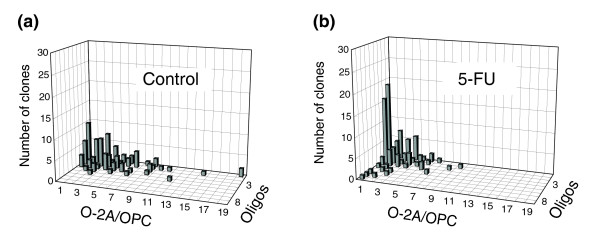
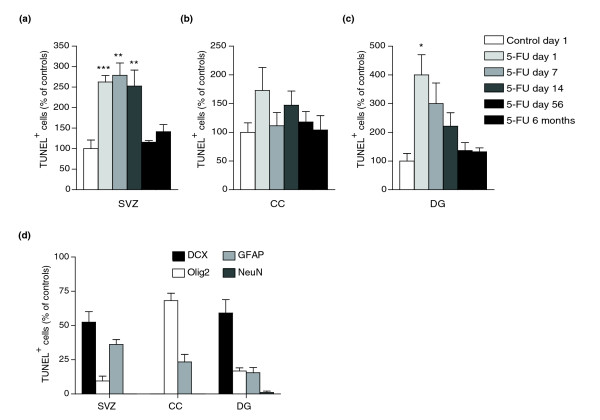
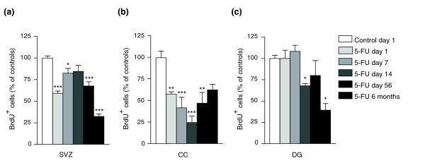
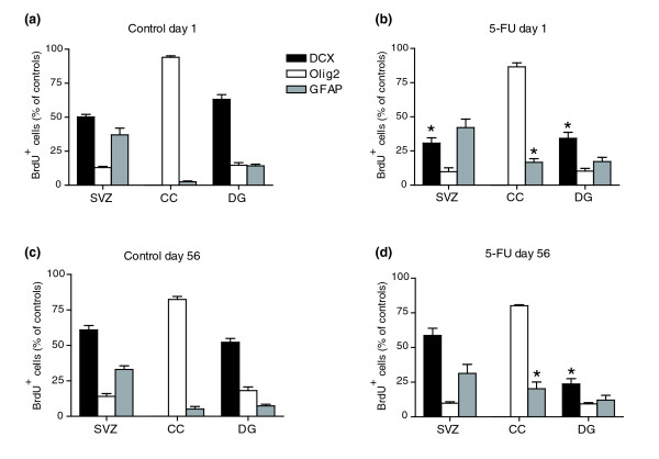
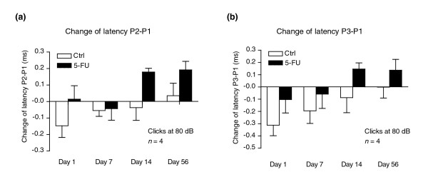
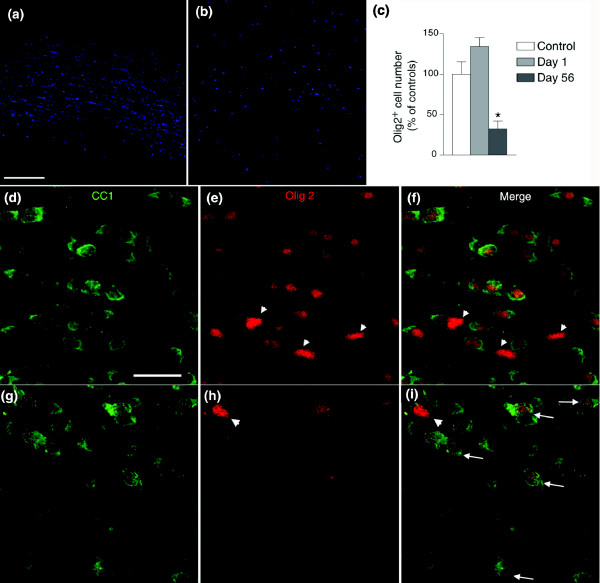
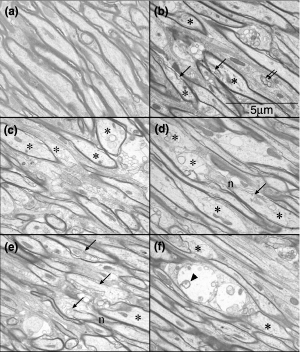
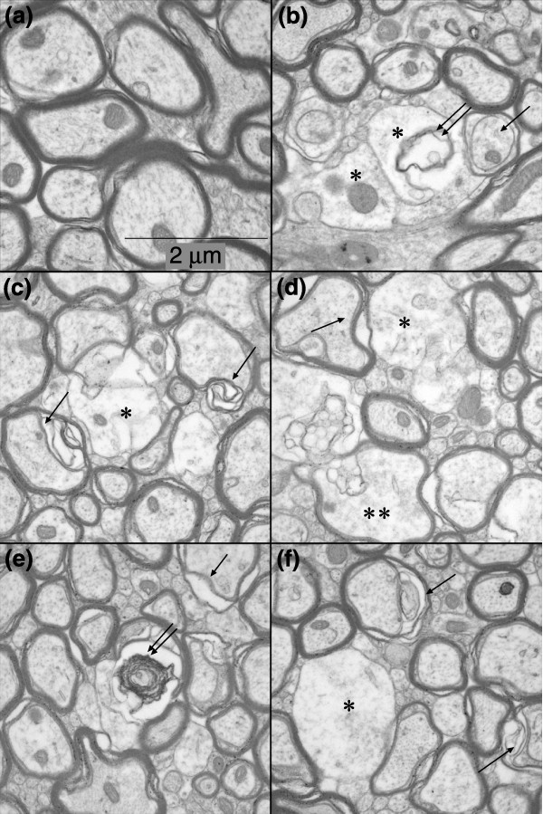
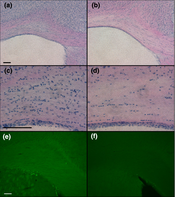
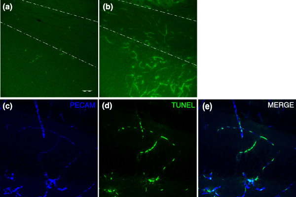
Results: We found that clinically relevant concentrations of 5-fluorouracil (5-FU; a widely used chemotherapeutic agent) were toxic for both central nervous system (CNS) progenitor cells and non-dividing oligodendrocytes in vitro and in vivo. Short-term systemic administration of 5-FU caused both acute CNS damage and a syndrome of progressively worsening delayed damage to myelinated tracts of the CNS associated with altered transcriptional regulation in oligodendrocytes and extensive myelin pathology. Functional analysis also provided the first demonstration of delayed effects of chemotherapy on the latency of impulse conduction in the auditory system, offering the possibility of non-invasive analysis of myelin damage associated with cancer treatment.
Conclusions: Our studies demonstrate that systemic treatment with a single chemotherapeutic agent, 5-FU, is sufficient to cause a syndrome of delayed CNS damage and provide the first animal model of delayed damage to white-matter tracts of individuals treated with systemic chemotherapy. Unlike that caused by local irradiation, the degeneration caused by 5-FU treatment did not correlate with either chronic inflammation or extensive vascular damage and appears to represent a new class of delayed degenerative damage in the CNS.
Figures
References
-
- Inagaki M, Yoshikawa E, Matsuoka Y, Sugawara Y, Nakano T, Akechi T, Wada N, Imoto S, Murakami K, Uchitomi Y. Smaller regional volumes of brain gray and white matter demonstrated in breast cancer survivors exposed to adjuvant chemotherapy. Cancer. 2007;109:146–156. doi: 10.1002/cncr.22368. - DOI - PubMed
-
- Silverman DH, Dy CJ, Castellon SA, Lai J, Pio BS, Abraham L, Waddell K, Petersen L, Phelps ME, Ganz PA. Altered frontocortical, cerebellar, and basal ganglia activity in adjuvant-treated breast cancer survivors 5–10 years after chemotherapy. Breast Cancer Res Treat. 2007;103:303–311. doi: 10.1007/s10549-006-9380-z. - DOI - PubMed
-
- Hurria A, Rosen C, Hudis C, Zuckerman E, Panageas KS, Lachs MS, Witmer M, van Gorp WG, Fornier M, D'Andrea G, Moasser M, Dang C, Van Poznak C, Hurria A, Holland J. Cognitive function of older patients receiving adjuvant chemotherapy for breast cancer: a pilot prospective longitudinal study. J Am Geriatr Soc. 2006;54:925–931. doi: 10.1111/j.1532-5415.2006.00732.x. - DOI - PubMed
Publication types
MeSH terms
Substances
Grants and funding
LinkOut - more resources
Full Text Sources
Other Literature Sources
