

Frequency and cause of disagreements in diagnoses for fetuses referred for ventriculomegaly
- PMID: 18430880
- PMCID: PMC5410935
- DOI: 10.1148/radiol.2472071067
Frequency and cause of disagreements in diagnoses for fetuses referred for ventriculomegaly
Abstract
Purpose: To prospectively assess the frequency and cause of disagreements in diagnoses at ultrasonography (US) and magnetic resonance (MR) imaging for fetuses referred for ventriculomegaly (VM).
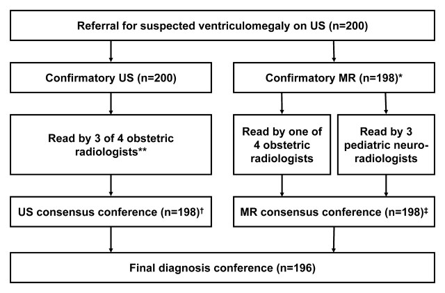
Materials and methods: One hundred ninety-five women, aged 18-44 years, with 200 fetal referrals for VM, were recruited in a prospective IRB-approved, HIPAA-compliant study. Written informed consent was obtained. US scans were prospectively interpreted by three obstetric radiologists and MR examinations were read by one obstetric radiologist and three pediatric neuroradiologists. Final diagnosis was reached by consensus (198 US, 198 MR, and 196 US-MR comparisons). Gestational age, ventricular size, types of disagreements, and reasons for disagreements were recorded. Interreader agreement was assessed with kappa statistics. Ventricular diameter, gestational age, and confidence scores were analyzed by using mixed-model analysis of variance, accounting for correlation within reader and fetus.
Results: There was prospective agreement on 118 (60%) of 198 US and 104 (53%) of 198 MR readings. Consensus was more likely when the final diagnosis was isolated VM (83 of 104, 80% at US; 82 of 109, 75% at MR) than when the final diagnosis included other anomalies as well (14 of 63, 22% at US; seven of 68, 10% at MR; P < .001). There was disagreement on 19 (10%) of 196 and 31 (16%) of 196 fetuses about the presence of VM at US and MR, respectively, and on 29 (15%) of 198 and 39 (20%) of 198 fetuses regarding the presence of major findings at US and MR, respectively. Reasons for discrepancies in reporting major findings included errors of observation, lack of real-time US scanning, lack of neuroradiology experience, as well as modality differences in helping depict abnormalities.
Conclusion: Of radiologists who read high-risk obstetric US and fetal MR images for VM, there is considerable variability in central nervous system diagnosis.
(c) RSNA, 2008.
Figures
Similar articles
-
Outcome of fetuses with cerebral ventriculomegaly and septum pellucidum leaflet abnormalities.AJR Am J Roentgenol. 2011 Jan;196(1):W83-92. doi: 10.2214/AJR.10.4434. AJR Am J Roentgenol. 2011. PMID: 21178039 Free PMC article.
-
Frequency and cause of disagreements in imaging diagnosis in children with ventriculomegaly diagnosed prenatally.Ultrasound Obstet Gynecol. 2010 Nov;36(5):582-95. doi: 10.1002/uog.7680. Ultrasound Obstet Gynecol. 2010. PMID: 20499405 Free PMC article.
-
MR imaging appearance of fetal cerebral ventricular morphology.Radiology. 2002 Jun;223(3):652-60. doi: 10.1148/radiol.2233011336. Radiology. 2002. PMID: 12034931
-
Role of magnetic resonance imaging in fetuses with mild or moderate ventriculomegaly in the era of fetal neurosonography: systematic review and meta-analysis.Ultrasound Obstet Gynecol. 2019 Aug;54(2):164-171. doi: 10.1002/uog.20197. Epub 2019 Jul 11. Ultrasound Obstet Gynecol. 2019. PMID: 30549340
-
Imaging of fetal cerebral ventriculomegaly: a guide to management and outcome.Semin Fetal Neonatal Med. 2005 Oct;10(5):421-8. doi: 10.1016/j.siny.2005.05.002. Semin Fetal Neonatal Med. 2005. PMID: 15985390 Review.
Cited by
-
Prevalence of defined ultrasound findings of unknown significance at the second trimester fetal anomaly scan and their association with adverse pregnancy outcomes: the Welsh study of mothers and babies population-based cohort.Prenat Diagn. 2016 Jan;36(1):40-8. doi: 10.1002/pd.4708. Epub 2015 Nov 20. Prenat Diagn. 2016. PMID: 26475362 Free PMC article. Clinical Trial.
-
Analysis of errors made on in utero MR studies of the foetal brain in the MERIDIAN study.Eur Radiol. 2019 Jan;29(1):195-201. doi: 10.1007/s00330-018-5508-x. Epub 2018 Jun 15. Eur Radiol. 2019. PMID: 29948083 Free PMC article.
-
Ultrasound and MRI of fetuses with ventriculomegaly: can cortical development be used to predict postnatal outcome?AJR Am J Roentgenol. 2011 Jun;196(6):1457-67. doi: 10.2214/AJR.10.5422. AJR Am J Roentgenol. 2011. PMID: 21606314 Free PMC article.
-
Multi-atlas multi-shape segmentation of fetal brain MRI for volumetric and morphometric analysis of ventriculomegaly.Neuroimage. 2012 Apr 15;60(3):1819-31. doi: 10.1016/j.neuroimage.2012.01.128. Epub 2012 Feb 10. Neuroimage. 2012. PMID: 22500924 Free PMC article.
-
Magnetic resonance volumetric assessments of brains in fetuses with ventriculomegaly correlated to outcomes.J Ultrasound Med. 2011 May;30(5):595-603. doi: 10.7863/jum.2011.30.5.595. J Ultrasound Med. 2011. PMID: 21527607 Free PMC article.
References
-
- Levine D, Barnes PD, Robertson RR, Wong G, Mehta TS. Fast MR imaging of fetal central nervous system abnormalities. Radiology 2003;229:51–61. - PubMed
-
- Levine D, Barnes PD, Madsen JR, Abbott J, Mehta T, Edelman RR. Central nervous system abnormalities assessed with prenatal magnetic resonance imaging. Obstet Gynecol 1999;94:1011–1019. - PubMed
-
- Poutamo J, Vanninen R, Partanen K, Ryynanen, Kirkinen P. Magnetic resonance imaging supplements ultrasonographic imaging of the posterior fossa, pharynx and neck in malformed fetuses. Ultrasound Obstet Gynecol 1999;13:327–334. - PubMed
-
- Dinh DH, Wright RM, Hanigan WC. The use of magnetic resonance imaging for the diagnosis of fetal intracranial anomalies. Childs Nerv Syst 1990;6:212–215. - PubMed
-
- Levine D, Barnes PD, Madsen JR, Li W, Edelman RR. Fetal central nervous system anomalies: MR imaging augments sonographic diagnosis. Radiology 1997;204:635–642. - PubMed
Publication types
MeSH terms
Grants and funding
LinkOut - more resources
Full Text Sources
Medical
