. 2008 Apr;178(4):1973-87.
doi: 10.1534/genetics.107.083469.

Genetic analysis of the Caenorhabditis elegans GLH family of P-granule proteins

Affiliations

Genetic analysis of the Caenorhabditis elegans GLH family of P-granule proteins

Caroline Spike et al. Genetics. 2008 Apr.

Abstract

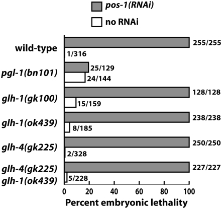
The Vasa DEAD-box helicases are widespread markers of germ cells across species, and in some organisms have been shown to be essential for germ-cell formation and development. In contrast to the single Vasa gene in most systems analyzed, Caenorhabditis elegans has four Vasa family members, the germline helicases GLH-1, GLH-2, GLH-3, and GLH-4. Our analysis of deletion alleles of each glh gene demonstrates that GLH-1 is the key member of the family: loss of GLH-1 function causes sterility that is mainly maternal effect, is manifested predominantly at elevated temperature, and is due to reduced germ-cell proliferation and impaired formation of both sperm and oocytes. The other GLHs are not essential. However, GLH-4 serves redundant roles with GLH-1: loss of both genes' function causes glh-1-like sterility at all temperatures. Molecular epistasis analysis demonstrates that GLH-1 and GLH-4 are required for proper association of the PGL family of proteins with P granules, suggesting a pathway of P-granule assembly in which the GLHs are upstream of the PGL proteins and the mRNA cap-binding protein IFE-1. While loss of some P-granule components causes worms to be defective in RNA interference, loss of GLH-1 and GLH-4 does not compromise RNAi. Thus, RNAi likely does not require intact P granules but instead relies on particular P-granule factors. We discuss the evolution of the Vasa/GLH genes and current views of their functions and the assembly and roles of germ granules among species.


Figures

F<sc>igure</sc> 1.—
Figure 1.—
Mutant alleles of glh-1. (A) Schematic of the glh-1 gene, showing the positions and molecular lesions of the four mutant alleles. Coding regions are shown as boxes and introns as lines. (B) Graph of the GLH-1 polypeptide predicted to be produced by each mutant. Box with light shading is a Gly-rich region. Solid box is a CCHC zinc finger. Dark shading is a DEAD-box helicase domain. The arrows in gk100 and ok439 indicate the positions of missing amino acids. The ok439 lesion is predicted to fuse a novel 10-amino-acid stretch to the carboxy terminus of GLH-1. The asterisk in bn103 shows the position of the Tc1 insertion. bn125 removes the last 28 amino acids. (C) Western blot analysis of the GLH-1 polypeptide produced by each mutant. The blot was reacted with affinity-purified rabbit anti-GLH-1 and mouse anti-actin as a loading control (shown by an asterisk). Values beneath the lanes are the levels of GLH-1, normalized to the actin level and expressed relative to wild type.
F<sc>igure</sc> 2.—
Figure 2.—
glh-1 mutations result in sterility that is sensitive to maternal genotype and temperature and is enhanced by a glh-4 mutation. (A) Analysis of zygotic (M+Z−) and maternal-effect (M−Z−) sterility at different temperatures. The number of worms analyzed ranged from 10 to >200. (B) Graph showing the generation of M+Z− and M−Z− worms. (C) Graphical display of sterility as a function of temperature for N2 (wild-type) and M−Z− glh-1, glh-4, and glh-4 glh-1 double mutants. glh-1 sterility is relatively low at 16°, 20°, and 24.5° and high at 26°. glh-4 glh-1 sterility is high at all temperatures.
F<sc>igure</sc> 3.—
Figure 3.—
glh-1 mutants display a range of germline sizes and gamete defects. glh/hT2∷gfp mothers were shifted from 20° to 26°. Homozygous glh/glh M−Z− hermaphrodites were DAPI stained ∼1 day beyond the L4 stage. Images represent a common phenotype for each strain. (A) Wild type. (B) glh-1(bn125) large germline. (C) glh-1(ok439) large germline containing endomitotically replicating oocytes (arrows). (D) glh-1(ok439) small germline. (E) glh-1(gk100) small germline. (F) glh-4(gk225) “Y”-shaped gonad observed in 1–15% of gonad arms. (G) glh-4(gk225) glh-1(ok439) very small germline. Bars for A–F and G, 50 μm.
F<sc>igure</sc> 4.—
Figure 4.—
Two mutant forms of GLH-1 are not detectably associated with P granules. glh M−Z− hermaphrodites derived from glh/hT2∷gfp grandmothers at 20° were cut, fixed, and stained with mouse PA3 to stain DNA (A, C, E, G, and I) and with rabbit anti-GLH-1 (B, D, F, H, and J) and imaged by confocal microscopy. Each panel shows a single ∼0.5-μm optical section in the pachytene region of the germline. (A and B) Wild type. (C and D) glh-1(gk100). (E and F) glh-1(ok439). (G and H) glh-1(bn125). (I and J) glh-4(gk225) glh-1(ok439). The alleles gk100 and ok439 cause dispersal of GLH-1 from P granules (D, F, and J). The bn125 allele causes only modest dispersal (H). Bar, 10 μm.
F<sc>igure</sc> 5.—
Figure 5.—
GLH-4 granules are not properly concentrated around nuclei in fixed glh-1 mutants. Worms were prepared as for Figure 4, but stained with mouse PA3 to stain DNA (A, C, and E) and with rabbit anti-GLH-4 (B, D, and F). Each panel shows a single ∼0.5-μm confocal section of the pachytene region of the germline. (A and B) Wild type. (C and D) glh-1(gk100). (E and F) glh-1(ok439). The alleles gk100 and ok439 cause some GLH-4 granules to lose their perinuclear localization (D and F). Bar, 10 μm.
F<sc>igure</sc> 6.—
Figure 6.—
PGL-1 is not properly concentrated on P granules in fixed glh-1 and glh-4 glh-1 mutants. Worms were prepared as for Figure 4, but stained with mouse PA3 to stain DNA (A, C, E, and G) and with rabbit anti-PGL-1 (B, D, F, and H). Each panel shows a single ∼0.5-μm confocal section of the distal tip plus pachytene region of the germline, and two panels include a 100- to 200-cell embryo as well. (A and B) Wild type. (C and D) glh-1(gk100). (E and F) glh-1(ok439). (G and H) glh-4(gk225) glh-1(ok439). The alleles gk100 and ok439 cause some PGL-1 to become dispersed in the cytoplasm, especially in the pachytene region (D and F). PGL-1 is completely dispersed in glh-4 glh-1 double mutants (H). Bar, 10 μm.
F<sc>igure</sc> 7.—
Figure 7.—
GFP∷PGL-1 is not properly concentrated on P granules in living glh-1 mutants. Live worms containing a GFP∷PGL-1 transgene were imaged by confocal microscopy. Images represent single ∼0.5-μm optical sections. (A and B) Wild type. (C and D) glh-1(bn103). (E and F) glh-1(bn125). The majority of GFP∷PGL-1 is dispersed in the cytoplasm of glh-1(bn103) embryos (C) and germlines (D). glh-1(bn125) worms display less dramatic dispersal of GFP∷PGL-1. Bars, 10 μm.
F<sc>igure</sc> 8.—
Figure 8.—
Mutant alleles of glh-2, glh-3, and glh-4. A schematic of each glh gene shows the position and extent of each deletion allele. Coding regions are shown as boxes and introns as lines.
F<sc>igure</sc> 9.—
Figure 9.—
glh-1 mutants and glh-4 glh-1 double mutants are not resistant to RNAi. Worms of the genotypes shown were fed bacteria expressing dsRNA against pos-1 and allowed to produce embryos at 20°. The following strains and generations were used: glh-1(ok439), glh-4(gk225), and pgl-1(bn101), M−Z− generation; glh-1(gk100) and glh-4(gk225) glh-1(ok439), M+Z− generation. pgl-1 mutants were resistant to pos-1 RNAi, as reported by Robert et al. (2005). The other strains were sensitive. glh-1(gk100) M−Z− animals also appear to be sensitive to pos-1(RNAi) (95% lethality), but glh-1(gk100) M−Z− animals not exposed to pos-1 dsRNA also laid a significant percentage of eggs that died (51%).

References

    1. Breitwieser, W., F. H. Markussen, H. Horstmann and A. Ephrussi, 1996. Oskar protein interaction with Vasa represents an essential step in polar granule assembly. Genes Dev. 10 2179–2188. - PubMed
    1. Brenner, S., 1974. The genetics of Caenorhabditis elegans. Genetics 77 71–94. - PMC - PubMed
    1. Carmichael, J. B., C. Stoica, H. Parker, J. M. McCaffery, A. J. Simmonds et al., 2006. RNA interference effector proteins localize to mobile cytoplasmic puncta in Schizosaccharomyces pombe. Traffic 7 1032–1044. - PubMed
    1. Chan, S. P., and F. J. Slack, 2006. MicroRNA-mediated silencing inside P-bodies. RNA Biol. 3 97–100. - PubMed
    1. Cheeks, R. J., J. C. Canman, W. N. Gabriel, N. Meyer, S. Strome et al., 2004. C. elegans PAR proteins function by mobilizing and stabilizing asymmetrically localized protein complexes. Curr. Biol. 14 851–862. - PubMed

Publication types

MeSH terms