. 2008 Apr;178(4):1989-2002.
doi: 10.1534/genetics.107.086298.

Raw mediates antagonism of AP-1 activity in Drosophila

Affiliations

Raw mediates antagonism of AP-1 activity in Drosophila

Katherine L Bates et al. Genetics. 2008 Apr.

Abstract

High baselines of transcription factor activities represent fundamental obstacles to regulated signaling. Here we show that in Drosophila, quenching of basal activator protein 1 (AP-1) transcription factor activity serves as a prerequisite to its tight spatial and temporal control by the JNK (Jun N-terminal kinase) signaling cascade. Our studies indicate that the novel raw gene product is required to limit AP-1 activity to leading edge epidermal cells during embryonic dorsal closure. In addition, we provide the first evidence that the epidermis has a Basket JNK-independent capacity to activate AP-1 targets and that raw function is required broadly throughout the epidermis to antagonize this activity. Finally, our mechanistic studies of the three dorsal-open group genes [raw, ribbon (rib), and puckered (puc)] indicate that these gene products provide at least two tiers of JNK/AP-1 regulation. In addition to Puckered phosphatase function in leading edge epidermal cells as a negative-feedback regulator of JNK signaling, the three dorsal-open group gene products (Raw, Ribbon, and Puckered) are required more broadly in the dorsolateral epidermis to quench a basal, signaling-independent activity of the AP-1 transcription factor.


Figures

F<sc>igure</sc> 1.—
Figure 1.—
raw mutants define an allelic series. (A, C, E, and G) Embryonic cuticles and (B, D, F, and H) dpp mRNA expression in stage 13 whole-mount embryos. (A) Cuticles derived from embryos homozygous for raw1, a null allele, exhibit significant dorsal closure and head involution defects, as well as an absence of ventral denticle belts (*); (B) in whole-mount embryos in situ, leading edge dpp expression extends to a depth of nine cells in the lateral epidermis (↓). (C) Cuticles derived from embryos homozygous for the raw2 allele, a strong hypomorph, also exhibit significant dorsal closure and head involution defects, as well as severely atrophied ventral denticle belts (*). (D) In whole-mount embryos in situ, dpp expression extends to a depth of seven cells in the lateral epidermis (↓). (E) Cuticles derived from embryos homozygous for the raw2418 allele, a hypomorph of intermediate strength, exhibit a strong dorsal pucker, in addition to hypertrophied ventral denticle belts (*). (F) In whole-mount embryos in situ, dpp expression extends to a depth of four cells in the lateral epidermis (↓). (G) Cuticles derived from embryos homozygous for rawlex1, a very weak hypomorph, exhibit a weak dorsal pucker and ventral denticle belts that are indistinguishable from wild type (*). (H) In whole-mount embryos in situ, dpp expression only minimally extends into the lateral epidermis, at most to a depth of two cells laterally (↓). In this and all subsequent figures, embryos are oriented such that dorsal is up and anterior is to the left.
F<sc>igure</sc> 2.—
Figure 2.—
Ventral denticle abnormalities in raw mutant embryos result from ectopic epidermal Dpp signaling. (A) Cuticles derived from raw1; UAS-brk/69BGAL4 embryos exhibit significant dorsal closure and head involution defects, comparable to those of the null homozygote shown in Figure 1A; the ventral denticle belt phenotype, however, is largely rescued. (B and C) Magnification and trace drawing of ventral denticles in A. brk mRNA transcript expression in situ is shown in stage 13 whole-mount wild-type embryos (D) and raw1 mutant embryos (E). Whereas brk is expressed in lateral and ventral stripes in wild-type embryos, these expression domains are missing in raw1 homozygotes.
F<sc>igure</sc> 3.—
Figure 3.—
raw-group genes exhibit an array of shared loss-of-function phenotypes. (A, E, and I) dpp mRNA transcript expression in whole mount embryos in situ; (B, F, and J) cuticular phenotypes; (C, G, and K) magnification of cuticular denticle phenotypes; and (D, H, and L) trace drawings of denticle magnifications. In wild-type embryos, dpp expression is limited to the single row of leading-edge epidermal cells (A), and the five ventral denticle rows are clearly evident (B–D). pucH246 homozygotes exhibit expanded dpp expression in the lateral epidermis (E), a dorsal cuticular hole (F), and hypertrophied ventral denticle belts that are similar to those in raw2418 hypomorphs (G and H). rib1 homozygotes exhibit expanded dpp expression as well as dorsal closure ( J) and ventral denticle defects (K and L).
F<sc>igure</sc> 4.—
Figure 4.—
raw and puc function independently to antagonize JNK/AP-1 signaling. (A) raw2418; pucE69 cuticles exhibit dorsal closure, head involution, and ventral denticle defects that define null alleles of raw (see also Figure 1A). (B) The pucE69 cuticular phenotype is more subtle; it is characterized by a dorsal pucker. (C) raw1; pucH246 cuticles exhibit a novel phenotype, which masks defects in dorsal closure diagnostic of cuticles derived from single-mutant null animals. (D) The rawlex1 rib1 cuticular phenotype is analogous to the rawlex1 (and raw1) null phenotype. Cuticle magnification is identical in all panels.
F<sc>igure</sc> 5.—
Figure 5.—
Raw is cytoplasmic. N-terminal Myc-tagged Raw (A–C) is cytoplasmic in COS-7 cells. (D–F) C-terminal FLAG-tagged Raw is cytoplasmic in COS-7 cells. (A) N-terminal Myc-tagged Raw was visualized using an anti-Myc antibody; (D) C-terminal FLAG-tagged Raw was visualized using an anti-FLAG. Nuclei were visualized by staining with DAPI (B and E). Merged images of fluorescent channels are shown (C and F).
F<sc>igure</sc> 6.—
Figure 6.—
raw-group genes share identical epistatic relationships with genes encoding JNK-signaling molecules. dpp mRNA transcript expression in whole-mount embryos in situ. (A) bsk1, (B) raw1 bsk1, (C) JraIA109; pucH246, (D) JraIA109; rib1, (E) bsk1; pucH246, and (F) bsk1; rib1. In A–F, the position of the leading edge is marked by an arrow (↓). (G and H) Diagrams showing alternative explanations for the epistatic relationships of JNK-signaling molecules to raw (and other raw-group genes) in leading-edge (LE) and epidermal (EP) cells more generally.
F<sc>igure</sc> 7.—
Figure 7.—
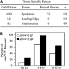
Raw functions early in the epidermis to effect dorsal closure. (A) Tissue-specific rescue in raw2418; p[UAS-raw+]/+ transgenics. Rescue was observed only when raw expression was driven by the 69B-GAL4 epidermal driver. In this experimental series, we observed rescue of embryonic dorsal closure in 72% of raw2418 homozygotes. (B) Induction of either of two independently derived hs:raw+ transgenes in raw2418 homozygotes 4–8 hr AEL rescues 68 and 98% of raw homozygous embryos to the first larval instar stage. Induction of the same transgenes 8–12 hr AEL rescues 39 and 65% of raw homozygotes. In the absence of heat shock, background rates of rescue range from 9 to 25%.
F<sc>igure</sc> 8.—
Figure 8.—
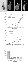
raw antagonizes JNK/AP-1 signaling at multiple Drosophila life stages. (A) raw mRNA transcripts are expressed in stretch cells; for this analysis oocytes were fixed and hybridized with an antisense RNA probe directed against raw. (B–D) raw-dependent gain- and loss-of-function egg phenotypes include size and dorsal appendage defects (indicated by arrows,↓). (B) +/+, (C) hs-raw+, and (D) raw1 follicle cell clones. (E and F) Quantization of gain- and loss-of-function-size phenotypes. (E) Eggs derived from wild-type females (dotted lines), exposed to either heat shock (♦) or not (⋄), are rarely smaller than 0.63 μm. In similar fashion, eggs derived from females harboring a raw+ transgene that has not been induced by heat shock (solid lines) are also only infrequently smaller than 0.63 μm (▵). In contrast, a significant fraction of eggs (34.8%) derived from transgenic females after raw+ induction by heat shock (▴). (F) Eggs derived from females harboring raw1 follicle cell clones exhibit a wider range of sizes than do controls. Twenty-one percent of eggs produced by FLP/FRT raw1 females (solid line) are smaller than 0.63 μm, whereas only 1.1% of eggshells produced by wild-type control females (dotted line) are smaller than 0.63 μm. (G) Reducing JNK/AP-1 signaling extends life span after exposure to oxidative stress. Wild-type adult females have an average lethality of 22.5% 24 hr after exposure to paraquat. Removal of one copy of puc reduces lethality to 2.0%. Removal of one copy of raw, using null (raw1 and rawlex1) or hypomorphic (raw2418) alleles reduces the lethality to 4.5 and 5.0%, respectively.
F<sc>igure</sc> 9.—
Figure 9.—
Modeling Raw function. (A) The canonical JNK cascade is active in LE cells and presumably controlled by the negative-feedback regulator Puckered. In the adjacent epidermal (EP) cells, Basket-independent AP-1 activity is silenced by Raw. (B) Baseline/basal levels of AP-1 activity are kept below biologically relevant levels by the actions of Raw, Ribbon, and Puckered. Activation of the JNK-signaling cascade in the leading-edge cells supersedes raw-group gene product-mediated AP-1 silencing. Lastly, biologically appropriate signaling levels are thought to be maintained via the activity of the JNK negative-feedback regulator Puckered.

References

    1. Arora, K., H. Dai, S. Kazuko, J. Jamal, M. O'Connor et al., 1995. The Drosophila schnurri gene acts in the Dpp/TGF beta signaling pathway and encodes a transcription factor homologous to the human MBP family. Cell 81 781–790. - PubMed
    1. Bishop, A. L., and A. Hall, 2000. Rho GTPases and their effector proteins. Biochem. J. 348(Pt 2): 241–255. - PMC - PubMed
    1. Blake, K. J., G. Myette and J. Jack, 1998. The products of ribbon and raw are necessary for proper cell shape and cellular localization of nonmuscle myosin in Drosophila. Dev. Biol. 203 177–188. - PubMed
    1. Blake, K. J., G. Myette and J. Jack, 1999. ribbon, raw, and zipper have distinct functions in reshaping the Drosophila cytoskeleton. Dev. Genes Evol. 209 555–559. - PubMed
    1. Bradley, P. L., and D. J. Andrew, 2001. ribbon encodes a novel BTB/POZ protein required for directed cell migration in Drosophila melanogaster. Development 128 3001–3015. - PubMed

Publication types

MeSH terms