

Stereomicroscopic fluorescence imaging of head and neck cancer xenografts targeting CD147
- PMID: 18431087
- PMCID: PMC2705783
- DOI: 10.4161/cbt.7.7.6109
Stereomicroscopic fluorescence imaging of head and neck cancer xenografts targeting CD147
Abstract
Purpose: To demonstrate that systemically administered fluorescently labeled anti-CD147 antibody can detect head and neck squamous cell carcinoma xenografts in vivo.
Experimental design: In vivo immunodeficient murine model.
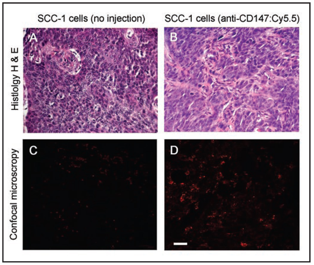
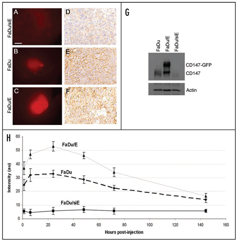
Results: Peak tumor fluorescence was visualized by near infrared stereomicroscopy in SCC-1 tumors at 24 hours after systemic injection of anti-CD147:Cy5.5 bioconjugate. SCC-1 xenografts demonstrated significantly higher fluorescent intensity after administration of CD147:Cy5.5 (48 au, p < 0.0001) compared to IgG1k:Cy5.5 isotype control antibody (9 au). FaDu tumors overexpressing CD147 (FaDu/E) demonstrated higher fluorescence (53 au) compared to control vector transfected cells (FaDu, 33 au, p < 0.0001) which was higher than CD147 knockdown cells (FaDu/siE, 5 au, p < 0.0001).
Methods: To determine if fluorescently labeled anti-CD147 antibody was specific for tumors in vivo, anti-CD147 and non-specific IgG1k antibody were labeled with a near infrared fluorophore (Cy5.5) and administered systemically to immunodeficient mice bearing SCC-1 xenografts. Imaging was performed over a 72 hour period using brightfield and fluorescent (685-735 nm) stereomicroscopy. To determine if fluorescence varied with receptor expression, SCID mice were xenografted with cell lines expressing variable amounts of CD147: FaDu (control vector transfected), FaDu/siE (siRNA CD147 knockdown) or FaDu/E (CD147 overexpressing) cells.
Conclusions: This data suggests fluorescently labeled anti-CD147 may have clinical utility in detection of HNSCC.
Figures


Comment in
-
CD147: a potential regulator of oncogenesis non-invasive imaging of CD147 in living subjects.Cancer Biol Ther. 2008 Jul;7(7):1071-2. doi: 10.4161/cbt.7.7.6617. Epub 2008 Jul 16. Cancer Biol Ther. 2008. PMID: 18698167 No abstract available.
References
-
- Cook JA, Jones AS, Phillips DE, Soler Lluch E. Implications of tumour in resection margins following surgical treatment of squamous cell carcinoma of the head and neck. Clin Otolaryngol Allied Sci. 1993;18:37–41. - PubMed
-
- Woolgar JA, Triantafyllou A. A histopathological appraisal of surgical margins in oral and oropharyngeal cancer resection specimens. Oral Oncol. 2005;41:1034–1043. - PubMed
-
- Ang KK, Trotti A, Brown BW, et al. Randomized trial addressing risk features and time factors of surgery plus radiotherapy in advanced head-and-neck cancer. International Journal of Radiation Oncology*Biology*Physics. 2001;51:571–578. - PubMed
-
- Lell M, Baum U, Greess H, et al. Head and neck tumors: imaging recurrent tumor and post-therapeutic changes with CT and MRI. European Journal of Radiology. 2000;33:239–247. - PubMed
-
- Hermans R, Pameijer FA, Mancuso AA, Parsons JT, Mendenhall WM. Laryngeal or Hypopharyngeal Squamous Cell Carcinoma: Can Follow-up CT after Definitive Radiation Therapy Be Used to Detect Local Failure Earlier than Clinical Examination Alone? Radiology. 2000;214:683–687. - PubMed
Publication types
MeSH terms
Substances
Grants and funding
LinkOut - more resources
Full Text Sources
Medical
Research Materials