Review
. 2008 May;67(5):377-87.
doi: 10.1097/NEN.0b013e31816ff780.

DNA damage and repair: relevance to mechanisms of neurodegeneration

Affiliations
Review

DNA damage and repair: relevance to mechanisms of neurodegeneration

Lee J Martin. J Neuropathol Exp Neurol. 2008 May.

Abstract

DNA damage is a form of cell stress and injury that has been implicated in the pathogenesis of many neurologic disorders, including amyotrophic lateral sclerosis, Alzheimer disease, Down syndrome, Parkinson disease, cerebral ischemia, and head trauma. However, most data reveal only associations, and the role for DNA damage in direct mechanisms of neurodegeneration is vague with respect to being a definitive upstream cause of neuron cell death, rather than a consequence of the degeneration. Although neurons seem inclined to develop DNA damage during oxidative stress, most of the existing work on DNA damage and repair mechanisms has been done in the context of cancer biology using cycling nonneuronal cells but not nondividing (i.e. postmitotic) neurons. Nevertheless, the identification of mutations in genes that encode proteins that function in DNA repair and DNA damage response in human hereditary DNA repair deficiency syndromes and ataxic disorders is establishing a mechanistic precedent that clearly links DNA damage and DNA repair abnormalities with progressive neurodegeneration. This review summarizes DNA damage and repair mechanisms and their potential relevance to the evolution of degeneration in postmitotic neurons.


Figures
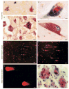
FIGURE 1
FIGURE 1
DNA damage and neuronal degeneration in human amyotrophic lateral sclerosis (ALS) and mouse models of motor neuron degeneration. (A) DNA damage in the form of 8-hydroxy-2-deoxyguanosine (OHdG) immunoreactivity (blue-green crystals) and p53 immunoreactivity (brown) colocalize in the nucleus of subsets of upper motor neurons (cell at right) in human ALS cerebral cortex. Other neurons (cell at left) have accumulated OHdG immunoreactivity within the cytoplasm, possibly corresponding to DNA damage within mitochondria, but are not positive for p53. Scale bar = 10 μm. (B) Activated p53, seen as phosphoserine392-p53 immunoreactivity (brown labeling), accumulates in subsets of cortical motor neurons (arrow) in human ALS. Other neurons in the field seen by the cresyl violet counterstain show no immunoreactivity. Scale bar = 10 μm. (C) In human ALS spinal motor neurons, nuclear OHdG-DNA damage (brown) and active caspase 3 (blue-green labeling) colocalize. Scale bar = 5 μm. (D) Mitochondrial accumulation identified by cytochrome c oxidase subunit 1 immunoreactivity (brown labeling) occurs in the perikaryon of human ALS spinal motor neurons with active caspase 3 (green in cytoplasm and nucleus). Many mitochondria are positive for active caspase 3. Scale bar = 5 μm. (E, F) Comet assay on isolated lumbar spinal cord motor neurons from G93A/mutant superoxide dismutase 1 mice at 6 (F) and 8 weeks (G) of age reveals the accumulation of DNA single-strand breaks (SSBs) in subsets of cells (arrows; see original publications for assay details; 19–21). Scale bars = (F) 60 and (G) 40 mm. (G, H) Spinal motor neurons with avulsed axons accumulate DNA-SSBs (detected by comet assay, note the short tails in G) very early in response to injury and coinciding with nuclear accumulation of p53 (brown nuclear labeling in H). Note that the p53 accumulation within the nucleus can be seen at chromatin strands. Scale bars = (G, H) 6 μm. mSOD-1, mutant superoxide dismutase 1.

References

    1. Lindahl T. Instability and decay of the primary structure of DNA. Nature. 1993;362:709–15. - PubMed
    1. Cleaver JE. Defective DNA repair replication in xeroderma pigmentosum. Nature. 1968;218:652–56. - PubMed
    1. Ronen A, Glickman BE. Human DNA repair genes. Environ Mol Mutagen. 2001;37:241–83. - PubMed
    1. Kraemer KH, Patronas NJ, Schiffmann R, et al. Xeroderma pigmentosum, trichothiodystrophy and Cockayne syndrome: A complex genotype-phenotype relationship. Neuroscience. 2007;145:1388–96. - PMC - PubMed
    1. Brooks PJ. DNA repair in neural cells: Basic science and clinical implications. Mutat Res. 2002;509:93–108. - PubMed

Publication types

Substances