. 2007 Oct;3(5):354-363.
doi: 10.1016/j.jpurol.2007.01.199.

Hypospadias and anorectal malformations mediated by Eph/ephrin signaling

Affiliations

Hypospadias and anorectal malformations mediated by Eph/ephrin signaling

Selcuk Yucel et al. J Pediatr Urol. 2007 Oct.

Abstract

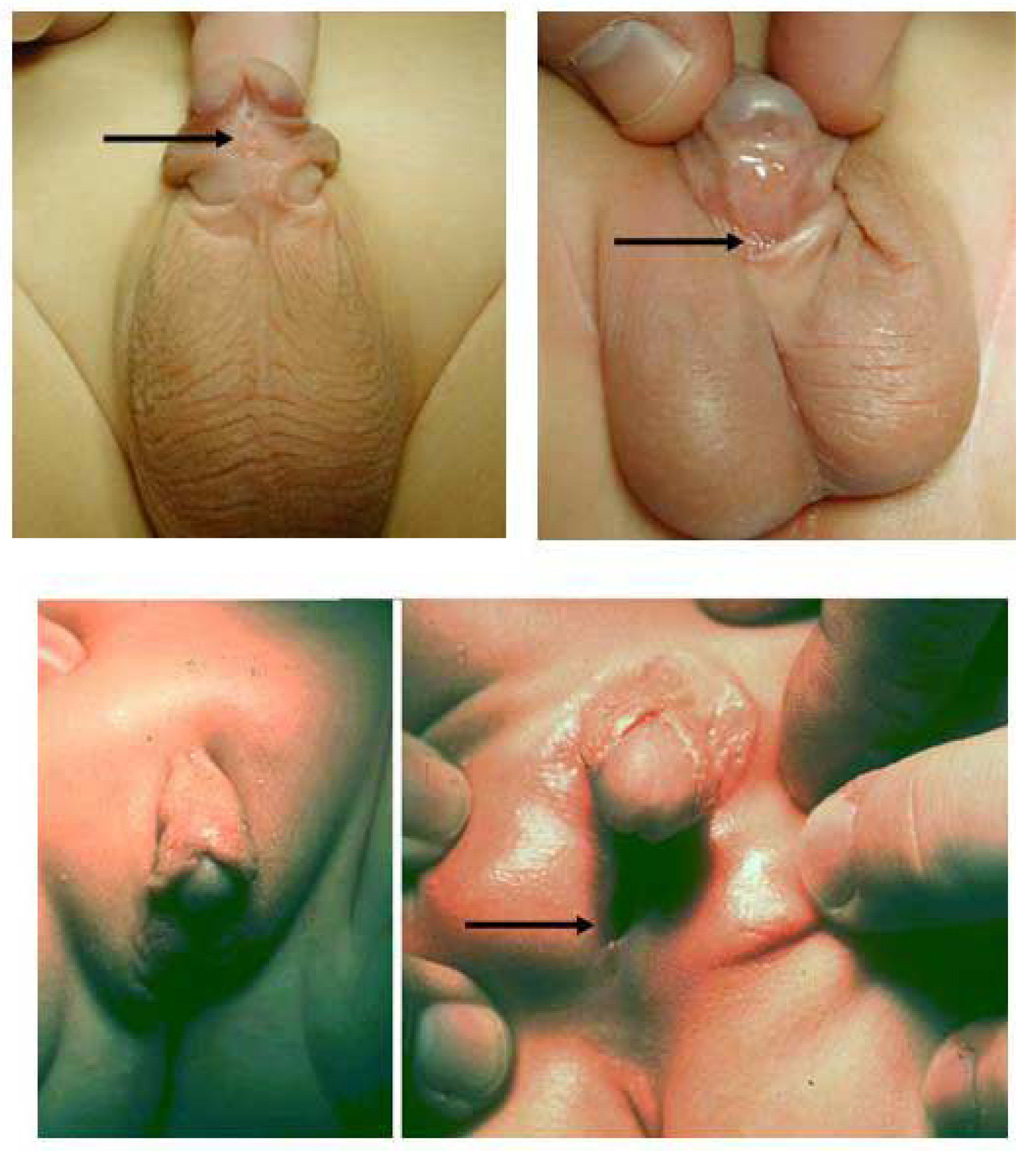
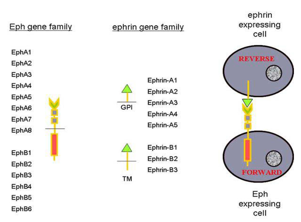
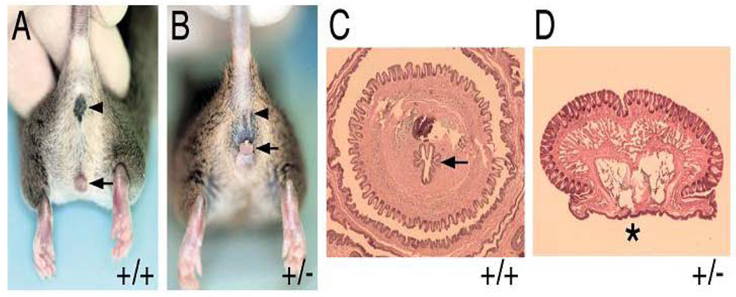
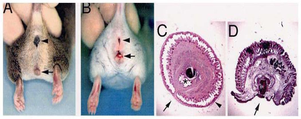
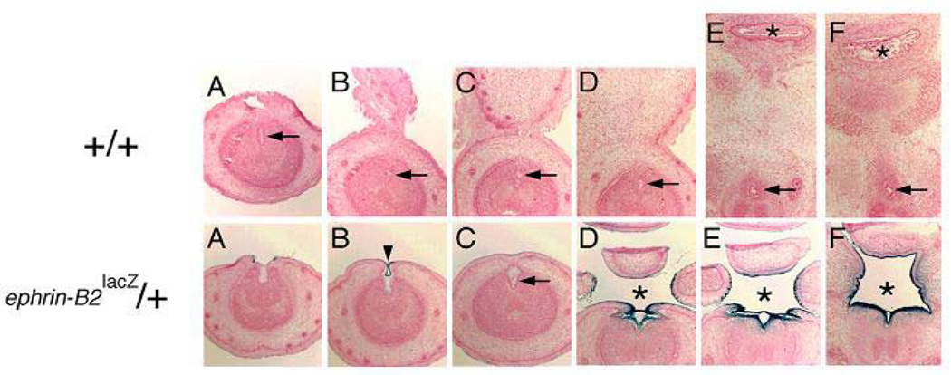
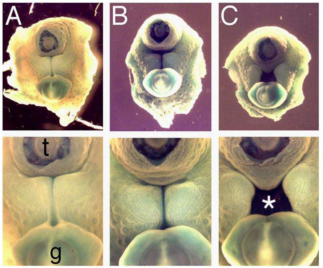
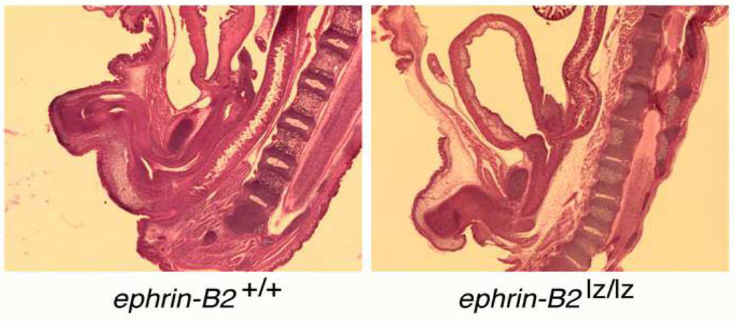
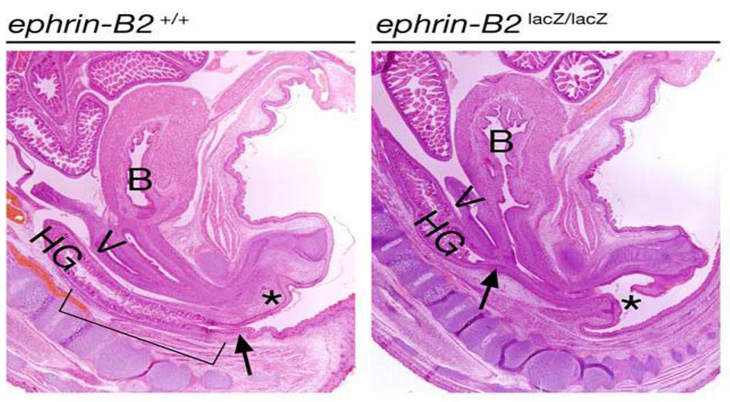
PURPOSE: Despite extensive research, the molecular basis of hypospadias and anorectal malformations is poorly understood, likely due to a multifactorial basis. The incidence of hypospadias is increasing, thus making research in this area warranted and timely. This review presents recent molecular work broadening our understanding of these disorders. MATERIALS AND METHODS: A brief review of our recent work and the literature on the role of Eph/ephrin signaling in hypospadias and anorectal malformations is presented. RESULTS: Genetically engineered mice mutant for ephrin-B2 or EphB2;EphB3 manifest a variety of genitourinary and anorectal malformations. Approximately 40% of adult male heterozygous mice demonstrate perineal hypospadias. Although homozygous mice die soon after birth, 100% of homozygous males demonstrate high imperforate anus with urethral anomalies and 100% of homozygous females demonstrate persistent cloaca. Male mice compound homozygous for EphB2(ki/ki);EphB3(Delta/Delta)/ also demonstrate hypospadias. CONCLUSIONS: These mouse models provide compelling evidence of the role of B-class Eph/ephrin signaling in genitourinary/anorectal development and add to our mechanistic and molecular understanding of normal and abnormal embryonic development. As research on the B-class Ephs and ephrins continues, they will likely be shown to be molecular contributors to the multifactorial basis of hypospadias and anorectal malformations in humans as well.


Figures

Figure 1
Figure 1
Three cases of human hypospadias demonstrating phenotypic variability. The arrow indicates the location of the urethral meatus in each case. (A) Subcoronal hypospadias. (B) Penoscrotal hypospadias with mild scrotal clefting. (C) Perineal hypospadias with ventral chordee and completely bifid scrotum.
Figure 2
Figure 2
Depiction of human embryonic development at weeks 5–7 of gestation. The allantois and hindgut drain into the cloaca. Classic embryology teaching suggests the cloaca is septated by the caudal and ventral growth of the Tourneux fold and the lateral to medial growth of the left and right Rathke folds. These folds coalesce, forming the urorectal septum which divides the primitive urogenital sinus from the anorectal canal. This process is controversial. Reproduced with permission.
Figure 3
Figure 3
(A) Male and female human external genitalia are identical at 6–7 weeks gestation as cloacal septation occurs. (B) Under the influence of androgens produced by the differentiating male testis, the genital tubercle elongates, the urethra tabularizes, the labioscrotal folds fuse, and the male perineum forms. Female external genitalia form in the absence of androgen action. Reproduced with permission.
Figure 4
Figure 4
The Eph and ephrin gene families are divided into A and B classes. Protein functional motifs are depicted by shapes; A-class ephrins are bound to the cell membrane by glycosylphosphotidy linositol (GPI) linkage while the B-class ephrins are transmembrane (TM) molecules. On the right, two cells are depicted, an Eph-expressing cell and an ephrin-expressing cell. Activation of the Eph receptor by an ephrin triggers the ‘forward’ signal in the Eph-expressing cell. Simultaneously, the ephrin is activated by the Eph, triggering the ‘reverse’ signal in the ephrin-expressing cell. Thus bidirectional signaling occurs and both gene families act as both receptors and ligands.
Figure 5
Figure 5
(A) Adult male wild-type (+/+) mouse depicting normal anus (arrowhead) and penis (arrow). (B) Adult male ephrin-B2lacZ/+ heterozygous(+/−) mouse with normal anus (arrowhead), reduced anogenital distance and perineal hypospadias (arrow). (C) Cross section of adult +/+ penis with tubularized urethra (arrow). (D) Cross section of adult-B2lacZ/+ heterozygous (+/−) mouse penis with hypospadias (asterisk). Reproduced with permission.
Figure 6
Figure 6
(A) Adult male wild-type (+/+) mouse depicting normal anus (arrowhead) and penis (arrow). (B) Adult male EphB2ki/ki;EphB3Δ/Δ compound homozygote mouse with normal anus (arrowhead), reduced anogenital distance and perineal hypospadias (arrow). (C) Cross section of adult +/+ penis with tubularized urethra (asterisk). (D) Cross section of adult EphB2ki/ki;EphB3Δ/Δ compound homozygous mouse penis with hypospadias (arrow). Reproduced with permission.
Figure 7
Figure 7
Upper panel: consecutive cross sections of embryonic day 17 (E17) wild-type mouse penis demonstrating tubularized urethra (arrow) and normal septated anus (asterisk). From left to right, the sections progress from the distal penis inward into the perineum. Lower panel: consecutive cross sections of embryonic day 17 (E17) ephrin-B2lacZ/+ heterozygous mouse penis demonstrating incomplete proximal urethral tubularization and perineal closure (asterisk). In some more distal locations, urethral tubularization appeared more normal (arrow and arrowhead). The dark-blue staining indicates the high expression of ephrin-B2lacZ in the tubularizing urethral epithelium, septating epithelium of the cloaca and urogenital sinus (not shown). From left to right, the sections progress from the distal penis inward into the perineum. Reproduced with permission.
Figure 8
Figure 8
Low power (upper panel) and high power (lower panel) view of the perineum of three E16 littermates with the amputated tail (t) and genital tubercle (g) viewed at top and bottom of photos, respectively. (A) EphB2χi/+;EphB3Δ/Δ mouse demonstrates closed perineum and tubularized urethra. (B and C) Two examples of EphB2χi/χi;EphB3Δ/Δ mice with mild (B) and severe (C) delays in perineal closure and urethral tubularization, with cloaca (asterisk). Reproduced with permission.
Figure 9
Figure 9
Left: sagittal section of E18 male wild-type mouse demonstrating normal spine, anorectum and male urethra. Right: sagittal section of E18 ephrin-B2lacZ/+ male with normal spine and high imperforate anus with rectourethral fistula at the bladder neck. Anal dimple is present.
Figure 10
Figure 10
Left: sagittal section of E18 female wild-type mouse demonstrating normal spine, bladder (B), anorectum–hindgut (HG), vagina (V) and female urethra (asterisk). Right: sagittal section of E18 ephrin-B2lacZ/+ female with normal spine and high persistent cloaca (the arrow). Anal dimple is present. Reproduced with permission.
Figure 11
Figure 11
E12.5 female EphB2lacZ/+ heterozygous genital tubercles were harvested and cultured in media with or without DHT for 2 days. The female genital tubercles responded to DHT exposure with phallic enlargement, urethral tubularization distally and increasing EphB2 expression in the glans and urethral plate distal to closure.

References

    1. Paulozzi LJ, Erickson D, Jackson RJ. Hypospadias trends in two US surveillance systems. Pediatrics. 1997;100:831–834. - PubMed
    1. Wilson JD, Griffin JE, George FW, Leshin M. The endocrine control of male phenotypic development. Aust J Biol Sci. 1983;36:101–128. - PubMed
    1. Wilson JD, Griffin JE, Russell DW. Steroid 5 alpha-reductase 2 deficiency. Endocr Rev. 1993;14:577. - PubMed
    1. Holmes N, Miller W, Baskin L. Lack of defects in androgen production in children with hypospadias. J Clin Endocrinol Metab. 2004;89:2811–2816. - PubMed
    1. Paulozzi L. International trends in rates of hypospadias and cryptorchidism. Environ Health Perspect. 1999;107:297–302. - PMC - PubMed