. 2008 Apr 23;3(4):e2009.
doi: 10.1371/journal.pone.0002009.

Etoposide induces ATM-dependent mitochondrial biogenesis through AMPK activation

Affiliations

Etoposide induces ATM-dependent mitochondrial biogenesis through AMPK activation

Xuan Fu et al. PLoS One. .

Abstract

Background: DNA damage such as double-stranded DNA breaks (DSBs) has been reported to stimulate mitochondrial biogenesis. However, the underlying mechanism is poorly understood. The major player in response to DSBs is ATM (ataxia telangiectasia mutated). Upon sensing DSBs, ATM is activated through autophosphorylation and phosphorylates a number of substrates for DNA repair, cell cycle regulation and apoptosis. ATM has been reported to phosphorylate the alpha subunit of AMP-activated protein kinase (AMPK), which senses AMP/ATP ratio in cells, and can be activated by upstream kinases. Here we provide evidence for a novel role of ATM in mitochondrial biogenesis through AMPK activation in response to etoposide-induced DNA damage.

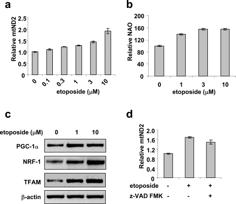
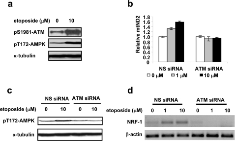
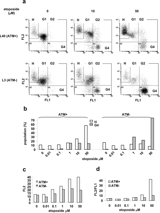
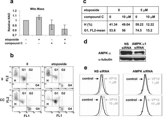
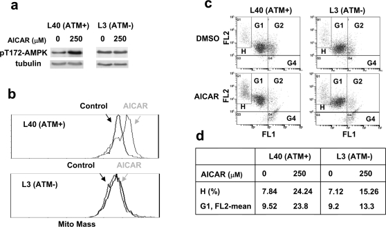
Methodology/principal findings: Three pairs of human ATM+ and ATM- cells were employed. Cells treated with etoposide exhibited an ATM-dependent increase in mitochondrial mass as measured by 10-N-Nonyl-Acridine Orange and MitoTracker Green FM staining, as well as an increase in mitochondrial DNA content. In addition, the expression of several known mitochondrial biogenesis regulators such as the major mitochondrial transcription factor NRF-1, PGC-1alpha and TFAM was also elevated in response to etoposide treatment as monitored by RT-PCR. Three pieces of evidence suggest that etoposide-induced mitochondrial biogenesis is due to ATM-dependent activation of AMPK. First, etoposide induced ATM-dependent phosphorylation of AMPK alpha subunit at Thr172, indicative of AMPK activation. Second, inhibition of AMPK blocked etoposide-induced mitochondrial biogenesis. Third, activation of AMPK by AICAR (an AMP analogue) stimulated mitochondrial biogenesis in an ATM-dependent manner, suggesting that ATM may be an upstream kinase of AMPK in the mitochondrial biogenesis pathway.

Conclusions/significance: These results suggest that activation of ATM by etoposide can lead to mitochondrial biogenesis through AMPK activation. We propose that ATM-dependent mitochondrial biogenesis may play a role in DNA damage response and ROS regulation, and that defect in ATM-dependent mitochondrial biogenesis could contribute to the manifestations of A-T disease.


Conflict of interest statement

Competing Interests: The authors have declared that no competing interests exist.

Figures

Figure 1
Figure 1. Etoposide induces mitochondrial biogenesis.
HeLa cells were treated with various concentrations of etoposide for 16 hrs. mtDNA content (a) and mitochondrial mass (b) were determined as described in the Materials and Methods. Data are presented as mean±SEM (n = 3). c. The mRNA levels of PGC1α, NRF−1 and TFAM were determined by RT-PCR. β-actin was used as a control for RT-PCR. d. HeLa cells were treated with 10 µM etoposide in the presence or absence of the caspase inhibitor z-VAD-FMK (20 µM) for 16 hrs and mtDNA content was then determined.
Figure 2
Figure 2. Etoposide-induced mitochondrial biogenesis is ATM-dependent.
a. Activation of ATM and AMPK were determined by Western blotting using specific antibodies against phosphorylated ATM at Ser1981 and phosphorylated AMPK at Thr172. α-tubulin was used as loading controls. HeLa cells were treated with 10 µM etoposide for 16 hrs. b–c. HeLa cells transfected with ATM siRNA or non-specific siRNA (NS siRNA) were treated with etoposide for 16 hrs. The mtDNA content was determined by quantitative real-time PCR (b). Phosphorylated AMPK at Thr172 was monitored by Western blotting (c). d. ATM siRNA and NS siRNA HeLa cells were treated with etoposide for 16 hrs. RNA was then isolated and the NRF-1 mRNA level was determined using RT-PCR.
Figure 3
Figure 3. ATM increases mitochondrial membrane potential in surviving population.
a. L40 (ATM+) and L3 (ATM−) cells were treated with indicated concentrations of etoposide for two days, followed by JC-1 staining and FACS analysis. Data were collected from normal living cell population that was gated according to the controls (no treatment) based on forward and side scatterings. b–d. Results were obtained from supplementary Fig. S4. b. Percentages of the H and the G4 populations in ATM+ and ATM− cells were plotted against etoposide concentrations. c. The mean intensities of FL2 in G1 population were plotted against etoposide concentrations. d. The ratios of the mean intensity of FL2 to the mean intensity of FL1 for G1 cells (including H cells) were plotted against etoposide. Data are a representative of three independent experiments.
Figure 4
Figure 4. Inhibition of AMPK abolishes etoposide-induced mitochondrial biogenesis.
a. Compound C blocks etoposide-induced increase in mitochondrial mass. L40 (ATM+) cells were treated with DMSO (control), 5 µM etoposide, 10 µM compound C, and 5 µM etoposide plus 10 µM compound C for 18 hrs. Cells were then stained by 1 µM NAO for FACS analysis. Data were obtained from three independent experiments. b–c. Compound C inhibits the increase in FL2 intensity of survival cells, as well as the increase of the H cell population. L40 (ATM+) cells were treated as described in a. Cells were then stained with JC-1 prior to FACS analysis. d. HeLa cells were transfected with siRNA against AMPK α or non-specific siRNA (NS). Knockdown of AMPK α was monitored by Western blotting using an antibody against AMPK α. e. AMPK α siRNA inhibits the increase in mitochondrial mass induced by etoposide. Cells were treated with indicated concentrations of etoposide (VP16) for 18 hrs. Cells were fixed by 60% ethanol and stained with MitoTracker Green FM prior to FACS analysis. 10,000 cells were analyzed.
Figure 5
Figure 5. AICAR induces ATM-dependent mitochondrial biogenesis.
a. Cells were treated with 250 µM AICAR for 100 min. Their lysates were then analyzed for activation of AMPK by using an antibody against phosphorylated AMPK α at Thr172. b. AICAR increases mitochondrial mass in ATM+, but not ATM− cells. Cells were treated with 250 µM AICAR for 2 days. They were then fixed and stained by 50 µM Mitotracker Green FM followed by FACS analysis. c–d. AICAR induces ATM-dependent increase in mitochondrial membrane potential. Cells were treated with 250 µM AICAR for 2 days, and then stained with JC-1, followed by FACS analysis.
Figure 6
Figure 6. Etoposide increases mitochondrial DNA content in mouse cortical neurons.
Isolated embryonic mouse cortical neurons (embryonic day 17.5) were treated with etoposide for 18 hrs. The relative amount of the mitochondrial mtND2 gene in mouse cortical neurons was measured by quantitative real-time PCR.
Figure 7
Figure 7. H2O2 treatment increases ATM autophosphorylation, and phosphorylations of p53, AMPK α and ACC (Acetyl-CoA Carboxylase).
HeLa cells were treated with indicated concentrations of H2O2 for 16 hrs followed by cell lysis. Western blottings were performed using antibodies against phosphorylated ATM (Ser1981), p53 (Ser15), AMPK α (Thr172) and ACC (Ser79), respectively.

References

    1. Taylor RW, Turnbull DM. Mitochondrial DNA mutations in human disease. Nat Rev Genet. 2005;6:389–402. - PMC - PubMed
    1. Zeviani M, Carelli V. Mitochondrial disorders. Curr Opin Neurol. 2003;16:585–594. - PubMed
    1. Sadun AA, Carelli V. Mitochondrial function and dysfunction within the optic nerve. Arch Ophthalmol. 2003;121:1342–1343. - PubMed
    1. Zeviani M, Spinazzola A, Carelli V. Nuclear genes in mitochondrial disorders. Curr Opin Genet Dev. 2003;13:262–270. - PubMed
    1. Scarpulla RC. Nuclear control of respiratory gene expression in mammalian cells. J Cell Biochem. 2006;97:673–683. - PubMed

Publication types

MeSH terms