. 2008 May;118(5):1815-24.
doi: 10.1172/JCI32467.

Ablation of GalNAc-4-sulfotransferase-1 enhances reproduction by altering the carbohydrate structures of luteinizing hormone in mice

Affiliations

Ablation of GalNAc-4-sulfotransferase-1 enhances reproduction by altering the carbohydrate structures of luteinizing hormone in mice

Yiling Mi et al. J Clin Invest. 2008 May.

Abstract

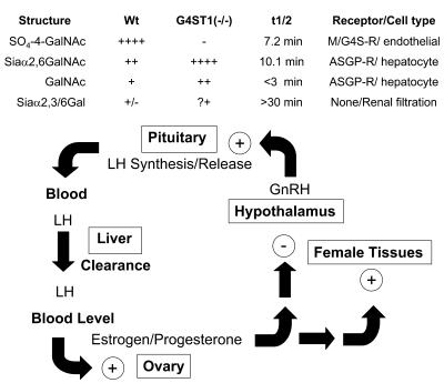
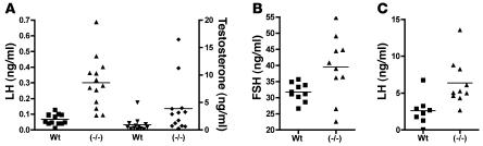
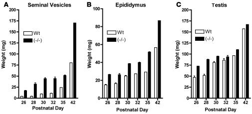
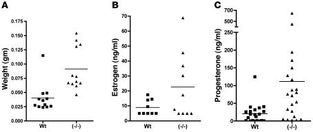
Luteinizing hormone (LH), produced in the anterior lobe of the pituitary, is a member of the hypothalamic-pituitary-gonad axis that is required for production of the sex hormones estradiol, progesterone, and testosterone. Perturbations in levels of hormones associated with this axis can result in defects in sexual development and maturity. LH bears unique N-linked carbohydrate units that terminate with a sulfated N-acetylgalactosamine structure (GalNAc-4-SO(4)) that mediates its clearance from the blood. To determine the significance of this terminal structure, we ablated the gene encoding the sulfotransferase responsible for sulfate addition to GalNAc on LH, GalNAc-4-sulfotransferase-1 (GalNAc-4-ST1) in mice. Mice lacking GalNAc-4-ST1 exhibited increased levels of circulating LH. In male mice, this resulted in elevated levels of testosterone and precocious maturation of testis and seminal vesicles. Female mice lacking GalNAc-4-ST1 demonstrated elevated estrogen levels and exhibited precocious sexual maturation and increased fecundity. Female mice remained in estrus for prolonged periods and produced almost 50% more litters per mouse than wild-type mice over the same period of time. Thus, sulfate modification of the terminal glycosylation of LH plays a central role in regulating the hypothalamic-pituitary-gonad axis in vivo.


Figures

Figure 1
Figure 1. Schematic of HPG axis.
The LH synthesized by WT mice bears N-linked oligosaccharides that terminate predominantly with SO4-4-GalNAc. Following GnRH-stimulated release into the blood, LH is rapidly removed from the blood by the Man/GalNAc-4-SO4-R expressed by endothelial cells in the liver, resulting in a half-life of 7.2 minutes in the blood. The amount of LH that reaches the LH receptor in the ovary and, as a result, the amount of estrogen/progesterone produced is determined by the half-life of LH. In the absence of GalNAc-4-4-ST1, the N-linked oligosaccharides on LH synthesized in the pituitary are predominantly modified with terminal Siaα2,6GalNAc. LH-bearing terminal Siaα2,6GalNAc is cleared by the ASGP-R expressed by hepatocytes, resulting in a longer half-life of 10.1 minutes. The longer half-life results in higher levels of LH in the blood and increased production of estrogen/progesterone by the ovary in GalNAc-4-ST1–/– mice as compared with WT mice. Thus, the rate of LH clearance and the concentration that LH attains in the blood are determined by the structures of its carbohydrate moieties. The strength of the signal produced by the same amount of LH released is determined by the structure of its terminal sugars. The impact of altered half-life appears to be superimposed on the other feedback mechanisms that normally regulate estrogen/progesterone levels. The same effects are seen in males with respect to T production.
Figure 2
Figure 2. Generation of GalNAc-4-ST1–/– mice.
(A) A targeting construct containing exons 3 and 4 of mouse GalNAc-4-ST1 flanked by loxP sites followed by PGK-neo also flanked by loxP sites was constructed as illustrated. 5′ and 3′ probes to regions outside of the area used to prepare the targeting construct were used to identify homologous recombinants in ES cells before and after treatment with Cre recombinase. (B) DNA from live progeny of heterozygous matings were genotyped by Southern blotting following EcoRI digestion. The WT product is 11.1 kb and the KO product is 10.3 kb. Examples of WT (+/–) and KO (–/–) progeny are shown.
Figure 3
Figure 3. LH, T, and FSH levels in WT and GalNAc-4-ST1–/– male mice.
Serum was collected from 8- to 10-week-old male WT and GalNAc-4-ST1–/– mice. Levels of LH, T, and FSH were determined by radioimmunoassay. In each case, the difference in mean values was significant. (A) P = 0.0001, LH (squares, triangles); P = 0.04, T (inverted triangles, diamonds). (B) P = 0.026, FSH. (C) P = 0.009, LH 5 days following castration.
Figure 4
Figure 4. Comparison of seminal vesicles and uteri from WT and GalNAc-4-ST1–/– mice.
(A) The seminal vesicles, prostate, and bladder were dissected from WT (left) and GalNAc-4-ST1–/– (right) male adult mice. The seminal vesicles are significantly enlarged. (B) The uterus and ovaries were dissected from WT (upper 2) and GalNAc-4-ST1–/– (lower 2) female adult mice. The uteri are enlarged in GalNAc-4-ST1–/– mice as compared with the WT mice. (C) Histologic sections of testis from postnatal day 26 and 28, WT and GalNAc-4-ST1–/–. Sperm can be seen in the section from GalNAc-4-ST1–/– mice on day 28 but not in the section from WT mice on day 28. Scale bars: 1 μm.
Figure 5
Figure 5. Comparison of weights for seminal vesicles, epididymides, and testes of WT and GalNAc-4-ST1–/– mice during postnatal development.
(A) Seminal vesicles, (B) epididymides, and (C) testes were carefully dissected from WT and GalNAc-4-ST1–/– male mice on the postnatal days indicated and weighed. Tissues were taken from 8, 18, 16, 14, 10, and 10 WT and 16, 19, 14, 22, 10, and 5 GalNAc-4-ST1–/– mice on postnatal days 26, 28, 30, 32, 35, and 42, respectively.
Figure 6
Figure 6. GalNAc-4-ST1–/– female mice display alterations in their estrus cycle and postnatal maturation.
Stages of the estrus cycle were determined for WT and GalNAc-4-ST1–/– mice by daily vaginal lavage for 19 consecutive days. (A) Examples of estrus cycles in WT and GalNAc-4-ST1–/– mice. P, proestrus; E, estrus; D1, diestrus1; D2, diestrus 2. (B) The number of cycles observed over 19 days was lower in 12 KO mice as compared with 9 WT mice; ***P < 0.0001. (C) Two groups of 15 WT and 15 GalNAc-4-ST1–/– female mice were examined daily beginning on postnatal day 25 for opening of the vagina, an indicator of having attained sexual maturity. The cumulative number of mice that attained maturity for each postnatal day is shown. WT mice, gray bars; KO mice, black bars.
Figure 7
Figure 7. Uterine weights, estrogen levels, and progesterone levels are increased in GalNAc-4-ST1–/– mice.
(A) The uteri from 10 WT and 10 GalNAc-4-ST1–/– adult female mice were collected and weighed. Means differed by 2.3-fold; P = 0.0007. The ovaries from WT and GalNAc-4-ST1–/– adult female mice were collected and extracts prepared by sonnication. (B) Estrogen level mean values differed by 2.5-fold; P = 0.0885. (C) Progesterone level mean values differed by 5.3-fold; P = 0.0118. The levels of estrogen and progesterone were determined by RIA.
Figure 8
Figure 8. GalNAc-4-ST1–/– mice do not add sulfate to N-linked carbohydrates on the glycoprotein hormones.
Extracts prepared from 5 WT and 5 GalNAc-4-ST1–/– mice that had not been castrated (A and C, left) or that had been castrated (B and C, right) were separated by SDS-PAGE and electrophoretically transferred to PVDF membranes for Western blot analysis. (A) Cys-Fc (chimeric protein specific for terminal GalNAc-4-SO4). (B) Mab6.3 (monoclonal antibody specific for SO4-4-GalNAcβ1,4GlcNAcβ1,2Man-). (C) Rabbit anti-LHβ. The location of the bands identified as the α subunit (not shown) and the LHβ subunit (C) in separate blots are indicated.
Figure 9
Figure 9. Clearance of endogenous LH in WT and GalNAc-4-ST1–/– mice.
WT and GalNAc-4-ST1–/– 36-week-old mice were surgically castrated or ovariectomized 5 days prior to the clearance studies. Serum was taken from the WT (n = 8) and GalNAc-4-ST1–/– (n = 8) castrated male mice and from the WT (n = 9) and GalNAc-4-ST1–/– (n = 6) ovariectomized female mice at the initiation of the clearance study. Acyline (10 μg/mouse) was administered intravenously, and blood was withdrawn at 5, 10, 20, and 30 minutes following introduction of acyline. (A) Comparison of LH clearance by individual castrated WT (squares) and castrated GalNAc-4-ST1–/– (triangles) mice following acyline injection. Half-lives for a total of 17 WT and 14 GalNAc-4-ST1–/– mice were determined using GraphPad Prism 4.0. (B) LH removed from the blood of WT mice had a half-life of 7.2 minutes (SEM = 0.6), and LH removed from the blood of GalNAc-4-ST1–/– mice had a half-life of 10.1 minutes (SEM = 0.9). The difference in half-lives is significant: *P = 0.01.
Figure 10
Figure 10. LH from GalNAc-4-ST1–/– mice has increased levels of Siaα2,6GalNAc.
Extracts were prepared from 5 WT and 5 GalNAc-4-ST1–/– mice that had not been castrated (A and B) or had been castrated (cast) (CF). Identical aliquots of each extract were incubated with immobilized WFA or SNA-1, which binds structures terminating with GalNAc or Siaα2,6GalNAc, respectively. Terminal sialic acid was removed by digestion with neuraminidase as indicated prior to incubation with WFA or SNA-1 (B, D, and F). Equal aliquots of the starting material (In), the unbound fraction (Ub), and the fractions selectively eluted with GalNAc or lactose (Elution) were analyzed by SDS-PAGE and Western blotting with anti-LHβ. (A) Binding of pituitary extracts by WFA-agarose. (B) Binding of pituitary extracts by WFA-agarose following neuraminidase digestion. (C) Binding of pituitary extracts from castrated mice by WFA-agarose. (D) Binding of pituitary extracts from castrated mice by WFA-agarose following neuraminidase digestion. (E) Binding by SNA-1–agarose. (F) Binding by SNA-1–agarose following digestion with neuraminidase.

References

    1. Ying S.Y. Inhibins, activins, and follistatins: gonadal proteins modulating the secretion of follicle-stimulating hormone. Endocr. Rev. 1988;9:267–293. - PubMed
    1. Strauss, J.F., 3rd, and Penning, T.M. 1999. Synthesis of the sex steroid hormones: molecular and structural biology with applications to clinical practise. InMolecular biology in reproductive medicine. B.C.J.M. Fauser, A.J. Rutherford, J.F. Strauss, 3rd, and A. Van Steirteghem, editors. The Parthenon Publishing Group. Carnforth, United Kingdom. 201–232.
    1. Bousfield, G.R., Perry, W.M., and Ward, D.N. 1994. Gonadotropins. Chemistry and biosynthesis. InThe physiology of reproduction. E. Knobil, and J.D. Neill, editors. Raven Press. New York, New York, USA. 1749–1792.
    1. Clarke I.J., Cummins J.T. The temporal relationship between gonadotropin releasing hormone (GnRH) and luteinizing hormone (LH) secretion in ovariectomized ewes. Endocrinology. 1982;111:1737–1739. - PubMed
    1. Belchetz P.E., Plant T.M., Nakai Y., Keogh E.J., Knobil E. Hypophysial responses to continuous and intermittent delivery of hypopthalamic gonadotropin-releasing hormone. Science. 1978;202:631–633. doi: 10.1126/science.100883. - DOI - PubMed

Publication types

MeSH terms