. 2008 May;118(5):1750-64.
doi: 10.1172/JCI34149.

Identification of kinetin riboside as a repressor of CCND1 and CCND2 with preclinical antimyeloma activity

Affiliations

Identification of kinetin riboside as a repressor of CCND1 and CCND2 with preclinical antimyeloma activity

Rodger E Tiedemann et al. J Clin Invest. 2008 May.

Abstract

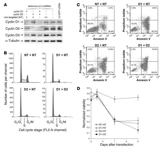
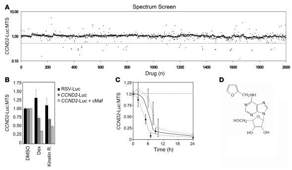
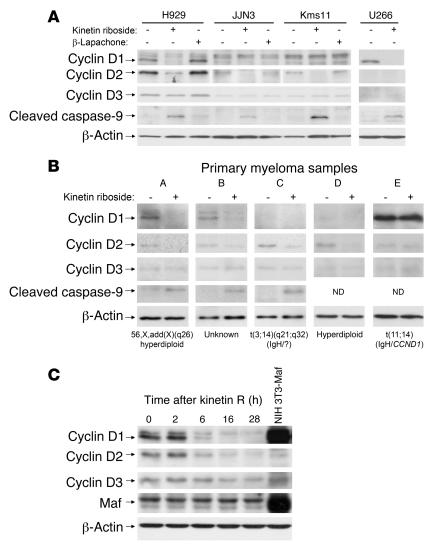
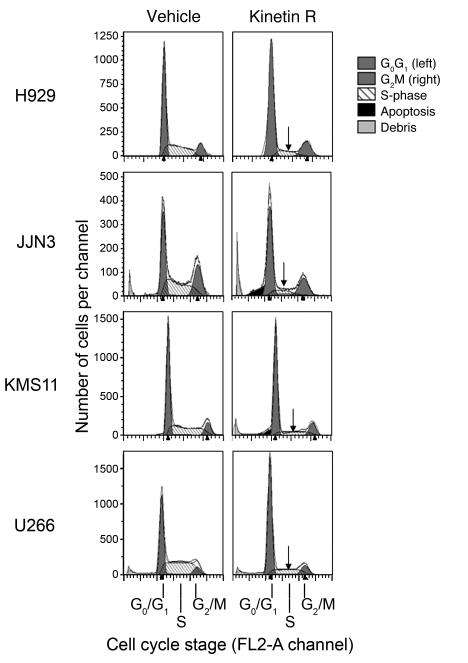
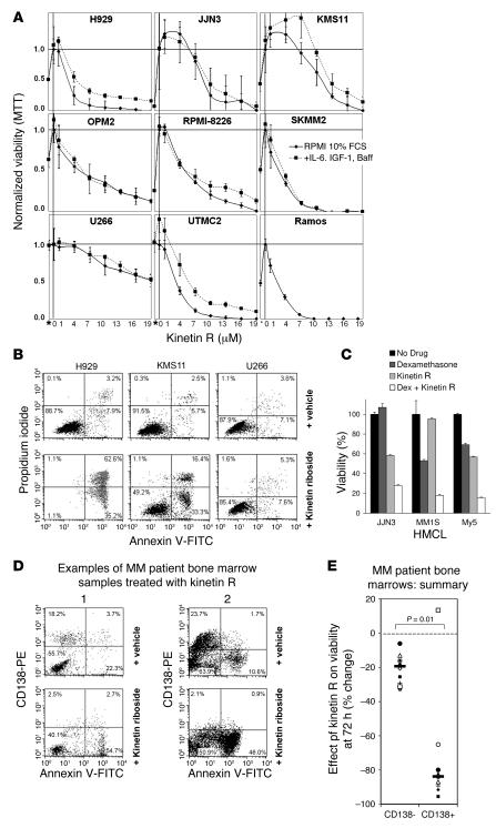
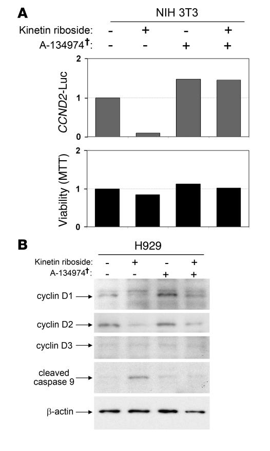
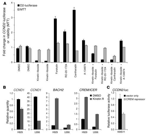
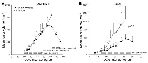
Knockout and transgenic studies in mice demonstrate that normal somatic tissues redundantly express 3 cyclin D proteins, whereas tumor cells seem dependent on a single overexpressed cyclin D. Thus, selective suppression of the individual cyclin D deregulated in a tumor represents a biologically valid approach to targeted cancer therapy. In multiple myeloma, overexpression of 1 of the cyclin D proteins is a ubiquitous feature, unifying at least 7 different initiating genetic events. We demonstrate here that RNAi of genes encoding cyclin D1 and cyclin D2 (CCND1 and CCND2, respectively) inhibits proliferation and is progressively cytotoxic in human myeloma cells. By screening a chemical library using a cell-based assay for inhibition of CCND2 trans-activation, we identified the plant cytokinin kinetin riboside as an inhibitor of CCND2 trans-activation. Kinetin riboside induced marked suppression of CCND2 transcription and rapidly suppressed cyclin D1 and D2 protein expression in primary myeloma cells and tumor lines, causing cell-cycle arrest, tumor cell-selective apoptosis, and inhibition of myeloma growth in xenografted mice. Mechanistically, kinetin riboside upregulated expression of transcription repressor isoforms of cAMP-response element modulator (CREM) and blocked both trans-activation of CCND2 by various myeloma oncogenes and cis-activation of translocated CCND1, suggesting induction of an overriding repressor activity that blocks multiple oncogenic pathways targeting cyclin D genes. These data support targeted repression of cyclin D genes as a therapeutic strategy for human malignancies.


Figures

Figure 1
Figure 1. Lentiviral shRNA suppression of cyclin D1 and D2 induces cell-cycle arrest and delayed apoptosis in H929 tumor cells.
(A) Immunoblot analysis of cyclin D1, D2, and D3 expression in H929 myeloma cells 65 hours after infection with lentiviruses expressing cyclin D1 or cyclin D2 shRNA, NT control shRNA, or GFP cDNA. α-Tubulin levels are shown as loading controls. (B) Cell-cycle profiles of H929 cultures 4 days after lentivirus infection, comparing the effects of suppression of cyclin D1 or D2 with control NT lentivirus infection and with lentivirus-induced suppression of both cyclin D1 and cyclin D2. To ensure equivalent exposure to lentivirus and shRNA, cells infected with both D1 lentivirus and D2 lentivirus (D1 + D2) are compared with cells infected with D1 lentivirus and NT lentivirus (D1 + NT), D2 lentivirus and NT lentivirus (D2 + NT), or 2 aliquots of NT lentivirus (NT + NT). (C) H929 apoptosis was examined serially by flow cytometry (in this example on day 11) after cyclin D suppression. Viable cells (left lower quadrants) are distinguished from early apoptotic (annexin V–positive) or late apoptotic (propidium iodide–positive) cells. (D) Time course of H929 viability after infection with D1 + NT, D2 + NT, D1 + D2, or control NT + NT lentiviruses. Viability was determined by annexin V and propidium iodide staining, as illustrated in C, and is normalized to uninfected H929; 5% error in viability is shown.
Figure 2
Figure 2. Identification of kinetin riboside by drug library screening for inhibitors of CCND2 promoter trans-activation in an NIH3T3 cell reporter system.
(A) Compounds were screened for inhibition of CCND2 promoter trans-activation at 16 hours in a 3T3 reporter model in which the MAF–trans-activating factor was coexpressed with the CCND2 promoter–driving LUC expression. The results of screening the Spectrum library is shown as a dot plot comparing each compound’s assay position (x axis) with its effect on MAF-driven CCND2 promoter trans-activation (measured by LUC) relative to effects on 3T3 viability (measured by MTS; y axis). Compounds below the dotted line were defined as putative hits. LOPAC and Prestwick drug libraries were also screened but are not shown. (B) Repeat testing of kinetin riboside (kinetin R), dexamethasone, and vehicle against reporter cells expressing LUC driven by a control RSV promoter or the CCND2 promoter, with and without MAF coexpression, showing that LUC suppression by these drugs is mediated specifically by the CCND2 promoter. Repression of CCND2 promoter activity by kinetin riboside is not restricted to trans-activation induced by MAF. (C) Suppression of CCND2-driven LUC protein levels in 3T3 following kinetin riboside (10 μM) treatment. Results of separate experiments are plotted (triangles and circles); each point is the mean of duplicates ± SEM. The overall curve of best fit is depicted as a solid line. Despite an estimated LUC half-life of 3 hours, kinetin riboside induces approximately 80% suppression by 9 hours, suggesting that suppression of the CCND2 promoter begins within 1–4 hours. (D) Chemical structure of kinetin riboside.
Figure 3
Figure 3. Kinetin riboside causes suppression of cyclins D1 and D2 and caspase activation in human MM.
(A) Immunoblot showing suppression of cyclins D1 and D2, but not D3, and induction of caspase-9 cleavage by kinetin riboside (10 μM) at 16 hours in HMCL. Kinetin riboside effects are compared with those of an unrelated control cytotoxic β-lapachone (1 μM, approximately 4 × IC50) or with DMSO vehicle. For visualization, the JJN3 cyclin D2 blot was exposed approximately 2-fold longer than cyclin D2 blots for H929, KMS11, and U266. (B) Kinetin riboside (10 μM) induces similar cyclin D1 and D2 suppression and caspase cleavage in CD138-purified primary myeloma cells at 16 hours, shown by immunoblotting. A plasma cell leukemia sample (patient E) was unaffected. The cytogenetics status of the tumor cells or IGH gene translocation is shown, if known. (C) Cyclin D and MAF protein levels in H929 cells after exposure to kinetin riboside (10 μM), showing suppression of cyclin D1 and D2 within 6 hours.
Figure 4
Figure 4. Kinetin riboside inhibits cell-cycle transit from G0/G1 to S-phase.
Cell-cycle profile of HMCL treated with DMSO vehicle or kinetin riboside (10 μM) at 20 hours. Left and right peaks represent G0/G1 and G2/M phases, respectively. Similar results were obtained with duplicate experiments.
Figure 5
Figure 5. Kinetin riboside induces growth arrest and apoptosis in HMCL and primary myeloma cells and shows synergistic cytotoxicity with corticosteroids.
(A) HMCL viability following kinetin riboside (1–20 μM) in the presence or absence of MM growth cytokines IL-6 (10 ng/ml), IGF-1 (100 ng/ml), and BAFF (25 ng/ml) by MTT assay at 72 hours. The Burkitt lymphoma line RAMOS is also shown. *MTT on day 0 without drug. (B) Induction of apoptosis in HMCL by vehicle or kinetin riboside at 96 hours, assessed by annexin V and propidium iodide uptake. (C) Synergistic cytotoxicity induced by kinetin riboside (5 μM) and dexamethasone (10 nM) in JJN3, MM1S, and MY5 HMCL, assessed by MTT assay at 48 hours; graph shows the mean of triplicates ± SEM. (D) Two examples of flow cytometric analyses of MM patient bone marrow samples treated with vehicle or kinetin riboside (10 μM), showing preferential loss of viable primary CD138+ tumor cells (left upper quadrants) versus CD138 hematopoietic cells (left lower quadrants) with kinetin riboside at 72 hours. Following kinetin riboside treatment, CD138+ plasma cells became annexin V positive and CD138 negative, consistent with apoptosis. (E) Effects of kinetin riboside on CD138+ and CD138 compartments in 10 MM patient bone marrow samples at 72 hours. CD138+ and CD138 viability responses are compared by Mann-Whitney test. CD138+ (primary tumor) cells are preferentially killed.
Figure 6
Figure 6. Cyclin D1 or D2 expressed from a heterologous CMV promoter is not suppressed by kinetin riboside and rescues KMS11 cells.
(A) Adenoviruses inducing expression of eGFP (control), cyclin D1 (CMV-D1), or cyclin D2 (CMV-D2) from heterologous CMV promoters were used to infect KMS11 or H929 cells; 48 hours later, cells were aliquotted and treated with DMSO vehicle (–) or kinetin riboside (15 μM) (+). Lysates were prepared at 12 hours; immunoblots show cyclin D protein levels following kinetin riboside treatment. Endogenously expressed cyclin D2 (lanes 1 and 3) and D1 (lane 9) is suppressed by kinetin riboside (lanes 2, 4, and 10), while CMV promoter–expressed cyclin D2 or D1 is not suppressed. (B) Cell-cycle studies on aliquots of cells from A were performed 20 hours after kinetin riboside treatment. The results are from 1 of 2 similar experiments. Expression of cyclin D1 or D2 from the heterologous CMV promoters rescues KMS11 cells from kinetin riboside–induced G0G1 cell-cycle arrest. (C) The proportion of KMS11 cells undergoing cell division following kinetin riboside treatment is summarized for cells expressing cyclin D1 or D2 from endogenous or CMV promoters, with an estimated 10% error. (D) Flow cytometry on adenovirus-infected KMS11 cells depicted in A 72 hours after treatment with vehicle or kinetin riboside (15 μM) showing infection status (eGFP expression) and viability (annexin V binding), comparing cells expressing cyclin D1 or D2 from endogenous or heterologous promoters. (E) Summary of kinetin riboside–induced apoptotic responses in KMS11 cells expressing cyclin D1 or D2 from endogenous or heterologous (CMV) promoters. Graph shows the measured percentage of viable (annexin V) virus-infected (GFP+) cells ± 10% error (estimated).
Figure 7
Figure 7. Kinetin riboside requires adenosine kinase for activity.
(A) Kinetin riboside–induced suppression of CCND2 promoter activation in 3T3 cells is abolished by the adenosine kinase inhibitor A-134974 (2 μM). 3T3 cells containing the CCND2 promoter–LUC reporter construct were preincubated with A-134974 (2 μM) for 1 hour and were then treated with kinetin riboside (10 μM) for 16 hours; CCND2 trans-activation and viability were determined by LUC and MTT assays, respectively. Specific adenosine kinase inhibitor. (B) Immunoblot of H929 MM cells showing that kinetin riboside–induced (10 μM) cyclin D suppression and caspase-9 cleavage are diminished by A-134974 (2 μM).
Figure 8
Figure 8. Kinetin riboside induces transcriptional repressors CREM and BACH2 and blocks cAMP- and PP2A-induced CCND2 promoter activation.
(A) 3T3 CCND2-LUC reporter cells were incubated with kinetin riboside or vehicle plus or minus forskolin, RO-20-1724, cantharidin, or A-134974, as specified (5 μM) for 16 hours; CCND2 trans-activation and viability were then assessed. Forskolin activates adenylate cyclase; RO-20-1724 inhibits cAMP phosphodiesterase; both cause increased cAMP signaling, which is seen to activate the CCND2 promoter. Cantharidin inhibits PP2A and also stimulates CCND2 promoter transcription. As shown, kinetin riboside blocks CCND2 promoter trans-activation by cAMP or PP2A-related phosphoproteins, initiated by forskolin, RO-20-1724, or cantharidin. Results represent 1 of 2 experiments and are shown as the mean of duplicate samples ± SEM. (B) H929 and U266 myeloma cells were treated with kinetin riboside (10 μM) for 4 hours and then processed for quantitative RT-PCR. Relative expression of genes determined by RT-PCR was normalized to H929 cells treated with vehicle alone and is plotted as the mean of replicates; error bars show the range. Consistent with gene-expression profiling analysis, quantitative RTPCR confirms that kinetin riboside rapidly induces CREM and BACH2 transcriptional repressors in both H929 and U266; parallel early suppression of CCND1 is also shown. (C) A pCMV-SPORT6 vector expressing the induced CREM repressor cDNA or a control pCMV-SPORT6 vector was cotransfected with the CCND2-LUC reporter into KMS11 myeloma cells, and CCND2 promoter activity was determined by LUC assay at 30 hours. Expression of the CREM repressor caused suppression of CCND2 promoter activity.
Figure 9
Figure 9. Kinetin riboside induces tumor growth arrest in MY5 and 8226 myeloma tumor xenografts.
(A) Matched pairs of nude mice bearing MY5 tumors were treated i.p. with vehicle control (open squares) or with kinetin riboside (filled squares) at a dose density escalating from 100 mg/kg once daily to 85 mg/kg 5×/daily, administered 5 days per week, commencing after mean tumor volume reached 135 mm3. Mean tumor volumes ± SEM are shown from the time of xenograft initiation (n = 4/group). (B) Nude mice bearing 8226 tumors were treated with vehicle (open squares) or with kinetin riboside (filled squares) (n = 4/group) commencing at 85 mg/kg 4 times daily (i.p. and s.c.) when the mean tumor volume exceeded 135 mm3. P value was calculated by paired t test.

Similar articles

Cited by

References

    1. [No authors listed]. 2004. Cancer facts and figures 2004. American Cancer Society. Atlanta, Georgia, USA. 58 pp.
    1. Bergsagel P.L., et al. Cyclin D dysregulation: an early and unifying pathogenic event in multiple myeloma. Blood. 2005;106:296–303. doi: 10.1182/blood-2005-01-0034. - DOI - PMC - PubMed
    1. Koziczak M., Holbro T., Hynes N.E. Blocking of FGFR signaling inhibits breast cancer cell proliferation through downregulation of D-type cyclins. Oncogene. 2004;23:3501–3508. doi: 10.1038/sj.onc.1207331. - DOI - PubMed
    1. Kozar K., et al. Mouse development and cell proliferation in the absence of D-cyclins. Cell. 2004;118:477–491. doi: 10.1016/j.cell.2004.07.025. - DOI - PubMed
    1. Malumbres M., et al. Mammalian cells cycle without the D-type cyclin-dependent kinases Cdk4 and Cdk6. Cell. 2004;118:493–504. doi: 10.1016/j.cell.2004.08.002. - DOI - PubMed

Publication types

MeSH terms