. 2008 May;118(5):1727-38.
doi: 10.1172/JCI34944.

STAT3 and STAT1 mediate IL-11-dependent and inflammation-associated gastric tumorigenesis in gp130 receptor mutant mice

Affiliations

STAT3 and STAT1 mediate IL-11-dependent and inflammation-associated gastric tumorigenesis in gp130 receptor mutant mice

Matthias Ernst et al. J Clin Invest. 2008 May.

Abstract

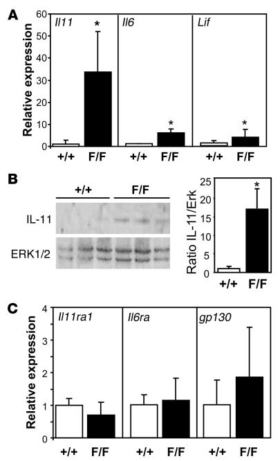
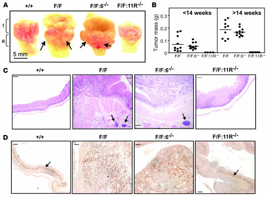
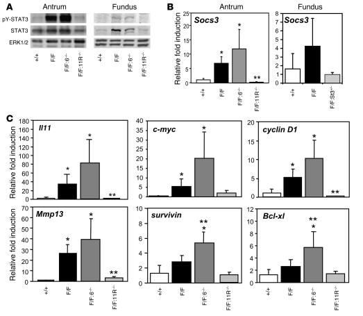
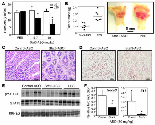
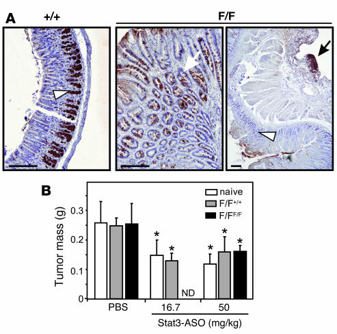
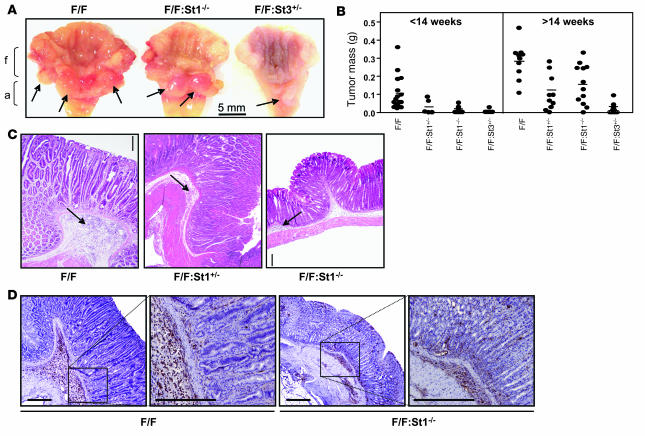
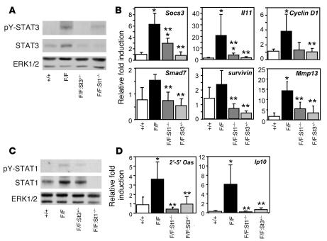
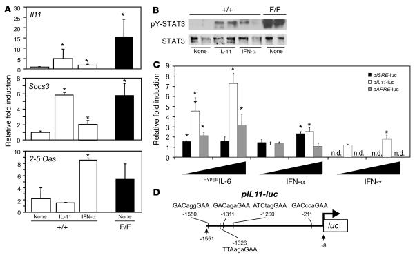
Deregulated activation of STAT3 is frequently associated with many human hematological and epithelial malignancies, including gastric cancer. While exaggerated STAT3 signaling facilitates an antiapoptotic, proangiogenic, and proproliferative environment for neoplastic cells, the molecular mechanisms leading to STAT3 hyperactivation remain poorly understood. Using the gp130(Y757F/Y757F) mouse model of gastric cancer, which carries a mutated gp130 cytokine receptor signaling subunit that cannot bind the negative regulator of cytokine signaling SOCS3 and is characterized by hyperactivation of the signaling molecules STAT1 and STAT3, we have provided genetic evidence that IL-11 promotes chronic gastric inflammation and associated tumorigenesis. Expression of IL-11 was increased in gastric tumors in gp130(Y757F/Y757F) mice, when compared with unaffected gastric tissue in wild-type mice, while gp130(Y757F/Y757F) mice lacking the IL-11 ligand-binding receptor subunit (IL-11Ralpha) showed normal gastric STAT3 activation and IL-11 expression and failed to develop gastric tumors. Furthermore, reducing STAT3 activity in gp130(Y757F/Y757F) mice, either genetically or by therapeutic administration of STAT3 antisense oligonucleotides, normalized gastric IL-11 expression and alleviated gastric tumor burden. Surprisingly, the genetic reduction of STAT1 expression also reduced gastric tumorigenesis in gp130(Y757F/Y757F) mice and coincided with reduced gastric inflammation and IL-11 expression. Collectively, our data have identified IL-11 as a crucial cytokine promoting chronic gastric inflammation and associated tumorigenesis mediated by excessive activation of STAT3 and STAT1.


Figures

Figure 1
Figure 1. Increased expression of IL-11 and other gp130 family cytokines in gastric tumors of gp130Y757F/Y757F mice.
(A) Quantitative RT-PCR (Q-PCR) analyses of Il11, Il6, and Lif gene expression were performed on cDNA derived from total RNA prepared from antral gastric tissue of 14-week-old gp130+/+ wild-type (+/+) and gp130Y757F/Y757F (F/F) mice. Expression data from 3–5 samples per genotype following normalization for 18S expression are shown and are presented from replicate analysis as the mean fold induction ± SD relative to expression observed in gp130+/+ samples. (B) Immunoblot analyses were performed on lysates prepared from antral gastric tissue of gp130+/+ and gp130Y757F/Y757F mice using the indicated antibodies. Each lane represents tissue from an individual mouse. Densitometric quantitation of IL-11 in each of 3 representative samples per genotype was performed and normalized against ERK1/2 protein levels. Data are presented as the mean fold induction ± SD relative to expression in gp130+/+ samples. (C) Q-PCR gene expression analyses of Il6ra, Il11ra1, and gp130 as in A. *P < 0.05 versus expression in gp130+/+ samples.
Figure 2
Figure 2. Complete abrogation of gastric tumor formation in gp130Y757F/Y757F mice lacking the IL-11Rα1 subunit.
(A) Representative appearance of stomachs from 14-week-old mice. Stomachs were opened along the outer curvature and pinned out with the lumen facing the viewer. Arrows indicate macroscopically visible lesions. Fundic (f) and antral (a) stomach regions are labeled. (B) Scatter plots depicting the total mass (g) of gastric tumors for individual mice either younger or older than 14 weeks of age and belonging to the indicated genotypes. Representative H&E- (C) and proliferating cell nuclear antigen–stained (D) cross-sections through the antral region of stomachs from mice of the indicated genotypes; arrows indicate patches of inflammatory cell accumulates (C) and the defined zone of proliferating mucous neck cells of the gastric epithelium (D). Scale bars: 100 μm. F/F: 11R–/– indicates gp130Y757F/Y757FIl11ra1–/– mice; F/F: 6–/– indicates gp130Y757F/Y757FIl6–/– mice.
Figure 3
Figure 3. Tumor suppression in gp130Y757F/Y757FIl11ra1–/– mice coincides with reduced IL-11 expression, STAT3 activation, and target gene expression.
(A) Immunoblot analyses were performed on lysates prepared from antral and fundic gastric tissues of 12- to 14-week-old mice using the indicated antibodies. Q-PCR expression analyses of Socs3 (B) in antral or fundic gastric tissue of the above mice, as well as Il11 and putative STAT3 target genes (C) were performed on cDNA derived from antral tissue of 14-week-old mice of the indicated genotypes. Expression data from 3–5 samples per genotype following normalization for 18S expression are shown and are presented from replicate analysis as the mean fold induction ± SD relative to expression in gp130+/+ samples. *P < 0.05 versus expression in gp130+/+ samples; **P < 0.05 versus expression in gp130Y757F/Y757F samples.
Figure 4
Figure 4. Reduced STAT3 activation and gastric tumor burden in STAT3-ASO–treated gp130Y757F/Y757F mice.
(A) Blood platelet counts of naive adult gp130Y757F/Y757F mice treated with either vehicle (PBS) or STAT3-ASO at the indicated concentrations every second day over 28 days. Data are expressed as mean ± SD. n = 8 for each treatment group. *P < 0.05 versus vehicle-treated mice. (B) Representative whole-mount appearance of stomachs from mice shown in A. Mice were treated either with vehicle or 50 mg/kg STAT3-ASO. Data points represent the combined wet weight of all excised polyps from individual mice, and the horizontal lines indicate the means. (C and D) Tissue cross-sections of gastric polyps from mice shown in B and stained with either H&E (C) or an antibody against BrdU (D). Scale bars: 100 μm. (E) Immunoblot analyses of antral gastric tissue lysates from individual naive gp130Y757F/Y757F mice using the indicated antibodies. Mice were treated with PBS, control-ASO, or STAT3-ASO at a final concentration of 50 mg/kg, and each lane represents an individual mouse. (F) Q-PCR analyses of Socs3 and Il11 gene expression in gastric tumors from mice shown in E and treated with either control ASO or STAT3-ASO at 50 mg/kg. Data are expressed as mean ± SD. n = 5 for each treatment group. *P < 0.05.
Figure 5
Figure 5. STAT3-dependent gastric tumorigenesis is independent of hematopoietic system–mediated antitumor immunity.
(A) Immunohistochemical analysis of pY-STAT3 in 12-week-old mice shows staining toward the base of the glandular gastric epithelium in wild-type and unaffected regions in gp130Y757F/Y757F mice (arrowheads), with extensive epithelial staining throughout the tumors (white arrow, middle panel) and of submucosal inflammatory cell infiltrates (black arrow, right panel). Scale bars: 100 μm. (B) The total wet weight of excised polyps from individual adult gp130Y757F/Y757F mice was determined following a 28-day treatment period with PBS vehicle or STAT3-ASO at the indicated concentration. Two months prior to ASO treatment, 6- to 8-week-old mice with established gastric tumors had been reconstituted with either autologous (F/FF/F) or heterologous gp130+/+ bone marrow (F/F+/+). Reconstituted mice were approximately 18–20 weeks old when sacrificed, and 12- to 14-week-old gp130Y757F/Y757F mice were included to control for potential radiation-mediated alterations to gastric polyp weights. Data are expressed as mean ± SD. n = 8 (naive mice), n = 5 (reconstituted mice). *P < 0.05 versus PBS-treated gp130Y757F/Y757F mice belonging to the corresponding group. ND, not determined.
Figure 6
Figure 6. Loss of STAT1 or STAT3 alleles in gp130Y757F/Y757F mice alleviates gastric tumor growth.
(A) Representative appearance of stomachs from 20-week-old mice belonging to the indicated genotypes. Arrows indicate macroscopically visible lesions in the antral but not the fundic regions of stomachs. (B) Scatter plots depicting total mass of gastric tumors from individual mice of the indicated genotypes and assessed in 2 different age groups. (C) Representative H&E-stained cross-sections through stomachs from 14-week-old mice of the indicated genotypes. (D) Representative immunohistochemical staining for CD45-positive hematopoietic cells in stomach sections from 14-week-old mice of the indicated genotypes. Scale bars in all histological sections: 100 μm. FF: St1+/– indicates gp130Y757F/Y757FStat1+/– mice.
Figure 7
Figure 7. Genetic restriction of STAT1 or STAT3 expression in gp130Y757F/Y757F mice coincides with reciprocal reduction in gastric STAT activation and associated target gene expression.
(A and C) Immunoblot analysis of antral gastric tissue lysates from 14-week-old mice of the indicated genotypes using antibodies specific for total ERK1/2, STAT1, or STAT3 and their corresponding tyrosine-phosphorylated forms. (B and D) Q-PCR analysis of gene expression on antral gastric tissue cDNA prepared from 14-week-old mice. Expression data from 3–5 mice per genotype following normalization for 18S expression are shown and are presented from replicate analysis as the mean fold induction ± SD relative to expression in gp130+/+ samples. *P < 0.05 versus expression in samples from gp130+/+ mice. **P < 0.05 versus expression in samples from gp130Y757F/Y757F mice.
Figure 8
Figure 8. STAT3 and STAT1 induce gastric IL-11 gene expression.
(A) Q-PCR on antral gastric cDNA prepared from untreated gp130Y757F/Y757F mice, as well as gp130+/+ mice either untreated or injected with a single dose of recombinant IL-11 or IFN-α. Expression data following normalization for 18S expression are shown and are presented from replicate analysis as the mean fold induction ± SD relative to expression in gp130+/+ samples. n = 3 mice per genotype and treatment. *P < 0.05 versus expression in untreated gp130+/+ samples. (B) Immunoblot analysis of total STAT3 and tyrosine-phosphorylated STAT3 in antral gastric tissue lysates from mice shown in A. (C) Transcriptional activation of the mouse Il11 gene in wild-type MEFs following transient transfection of the pIL11-luc firefly luciferase reporter construct. Triplicate cultures of cells, cotransfected with pTK-RL, were stimulated with the indicated amount of recombinant IFN-α, IFN-γ, or HYPERIL-6. Luciferase activity was determined 48 hours later and normalized against activity of the Renilla luciferase reporter and expressed relative to unstimulated control cultures. pAPRE-luc and pISRE-luc transfectants were used to assess for specific STAT3- and STAT1-mediated reporter activation. Data are representative of replicate analyses and are expressed as means of fold changes relative to unstimulated controls ± SD. *P < 0.05 versus expression in unstimulated cultures. (D) Schematic representation of pIL11-luc reporter plasmid depicting positions of potential STAT3 (top) and STAT1 (bottom) binding sites relative to the 5′ end of exon 1 of the mouse Il11 gene. The palindromic core motifs of the STAT binding sites are indicated in upper-case letters, and spacer sequences are indicated in lower-case letters.

Comment in

Similar articles

Cited by

References

    1. Parkin D.M., Bray F., Ferlay J., Pisani P. Global Cancer Statistics, 2002. CA Cancer J. Clin. . 2005;55:74–108. - PubMed
    1. Uemura N., et al. Helicobacter pylori infection and the development of gastric cancer. N. Engl. J. Med. 2001;345:784–789. doi: 10.1056/NEJMoa001999. - DOI - PubMed
    1. Wang J.Y., et al. Mutations of p53 gene in gastric carcinoma in Taiwan. Anticancer Res. . 2001;21:513–520. - PubMed
    1. Park W.S., et al. Somatic mutations of the trefoil factor family 1 gene in gastric cancer. Gastroenterology. 2000;119:691–698. doi: 10.1053/gast.2000.16483. - DOI - PubMed
    1. Guilford P., et al. E-cadherin germline mutations in familial gastric cancer. Nature. 1998;392:402–405. doi: 10.1038/32918. - DOI - PubMed

Publication types

MeSH terms