Review
. 2008 Sep;456(6):1005-24.
doi: 10.1007/s00424-008-0498-1. Epub 2008 Apr 23.

Physiology and pathophysiology of the vasopressin-regulated renal water reabsorption

Affiliations
Review

Physiology and pathophysiology of the vasopressin-regulated renal water reabsorption

Michelle Boone et al. Pflugers Arch. 2008 Sep.

Abstract

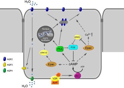
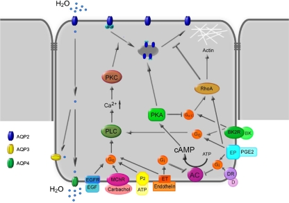
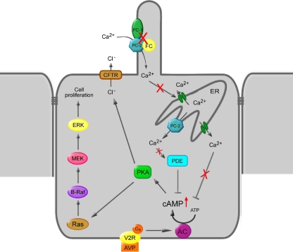
To prevent dehydration, terrestrial animals and humans have developed a sensitive and versatile system to maintain their water homeostasis. In states of hypernatremia or hypovolemia, the antidiuretic hormone vasopressin (AVP) is released from the pituitary and binds its type-2 receptor in renal principal cells. This triggers an intracellular cAMP signaling cascade, which phosphorylates aquaporin-2 (AQP2) and targets the channel to the apical plasma membrane. Driven by an osmotic gradient, pro-urinary water then passes the membrane through AQP2 and leaves the cell on the basolateral side via AQP3 and AQP4 water channels. When water homeostasis is restored, AVP levels decline, and AQP2 is internalized from the plasma membrane, leaving the plasma membrane watertight again. The action of AVP is counterbalanced by several hormones like prostaglandin E2, bradykinin, dopamine, endothelin-1, acetylcholine, epidermal growth factor, and purines. Moreover, AQP2 is strongly involved in the pathophysiology of disorders characterized by renal concentrating defects, as well as conditions associated with severe water retention. This review focuses on our recent increase in understanding of the molecular mechanisms underlying AVP-regulated renal water transport in both health and disease.


Figures

Fig. 1
Fig. 1
Model of the regulation of water permeability in renal collecting duct cells. Binding of vasopressin (AVP) to the V2 receptor (V2R) in the basolateral membrane activates adenylate cyclase (AC) and increases intracellular cAMP levels. This activates protein kinase A (PKA), which induces translocation of AQP2-bearing vesicles to the apical membrane, rendering this membrane water permeable. In addition, cAMP can activate exchange protein directly activated by cAMP (Epac). Epac can increase cytosolic Ca2+, which may facilitate AQP2 translocation. cAMP signaling is abrogated by phosphodiesterase (PDE)-mediated degradation of cAMP. Epac might control PDE activity by inhibiting ERK5, which inhibits PDE. PKA also increases AQP2 synthesis by phosphorylation of the cAMP-responsive element-binding (CREB) protein and its binding to the AQP2 promoter. Possibly, Epac enhances AQP2 synthesis by inhibiting extracellular signal-regulated kinases 1 and 2. Water, entering the principal cell via AQP2, can leave the cell via constitutively expressed AQP3 and AQP4
Fig. 2
Fig. 2
Model of the inhibition of AQP2-mediated water reabsorption. Several hormones can antagonize AVP-induced water transport. Indicated are AC adenylate cyclase, ATP adenosine tri-phosphate, BK(2R) bradykinin (type-2 receptor), cAMP cyclic adenosine monophosphate, D(R) dopamine (receptor), EGF(R) epidermal growth factor (receptor), EP E-prostanoid receptor, ET endothelin receptor, G α13 G protein involved in Rho family GTPase signaling, G i inhibitory G protein, G q PLC activating G protein, MChR muscarinic cholinergic receptor, P2 purinergic receptor, PGE2 prostaglandin-E2, PKA protein kinase A, PKC protein kinase C, PLC phospholipase C, Ub ubiquitin. For details, see text
Fig. 3
Fig. 3
Model of the pathways involved in polycystic kidney disease. Upon mechanical stimulation of the primary cilium, Ca2+ enters the cell via polycystin-2 (PC-2), which forms a complex with polycystin-1 (PC-1) and fibrocystin (FC). In addition, Ca2+ is released from the endoplasmatic reticulum (ER). Disruption of the polycystin/fibrocystin pathway results in decreased cytosolic Ca2+ levels. Reduced Ca2+ levels stimulate adenylate cyclase (AC) and inhibit phosphodiesterase (PDE), resulting in increased cAMP levels. Subsequent activation of protein kinase A (PKA) stimulates cell proliferation by sequential activation of Ras, B-Raf, MEK, and ERK. Furthermore, in polycystic kidney disease, cAMP-PKA signaling increases the permeability and expression of CFTR Cl channels, resulting in Cl extrusion. This increases the movement of Na and, subsequently, water into the lumen of the cyst

References

    1. Ahrabi AK, Terryn S, Valenti G, Caron N, Serradeil-Le GC, Raufaste D, Nielsen S, Horie S, Verbavatz JM, Devuyst O. PKD1 haploinsufficiency causes a syndrome of inappropriate antidiuresis in mice. J Am Soc Nephrol. 2007;18:1740–1753. - PubMed
    1. Alfie ME, Alim S, Mehta D, Shesely EG, Carretero OA. An enhanced effect of arginine vasopressin in bradykinin B2 receptor null mutant mice. Hypertension. 1999;33:1436–1440. - PubMed
    1. Ando Y, Asano Y. Functional evidence for an apical V1 receptor in rabbit cortical. Am J Physiol. 1993;264:F467–F471. - PubMed
    1. Ando Y, Tabei K, Asano Y. Luminal vasopressin modulates transport in the rabbit cortical collecting duct. J Clin Invest. 1991;88:952–959. - PMC - PubMed
    1. Aromataris EC, Roberts ML, Barritt GJ, Rychkov GY. Glucagon activates Ca2 and Cl− channels in rat hepatocytes. J Physiol. 2006;573:611–625. - PMC - PubMed

Publication types

LinkOut - more resources