. 2008 Jun 5;453(7196):807-11.
doi: 10.1038/nature06905. Epub 2008 Apr 23.

NF-kappaB links innate immunity to the hypoxic response through transcriptional regulation of HIF-1alpha

Affiliations

NF-kappaB links innate immunity to the hypoxic response through transcriptional regulation of HIF-1alpha

Jordi Rius et al. Nature. .

Abstract

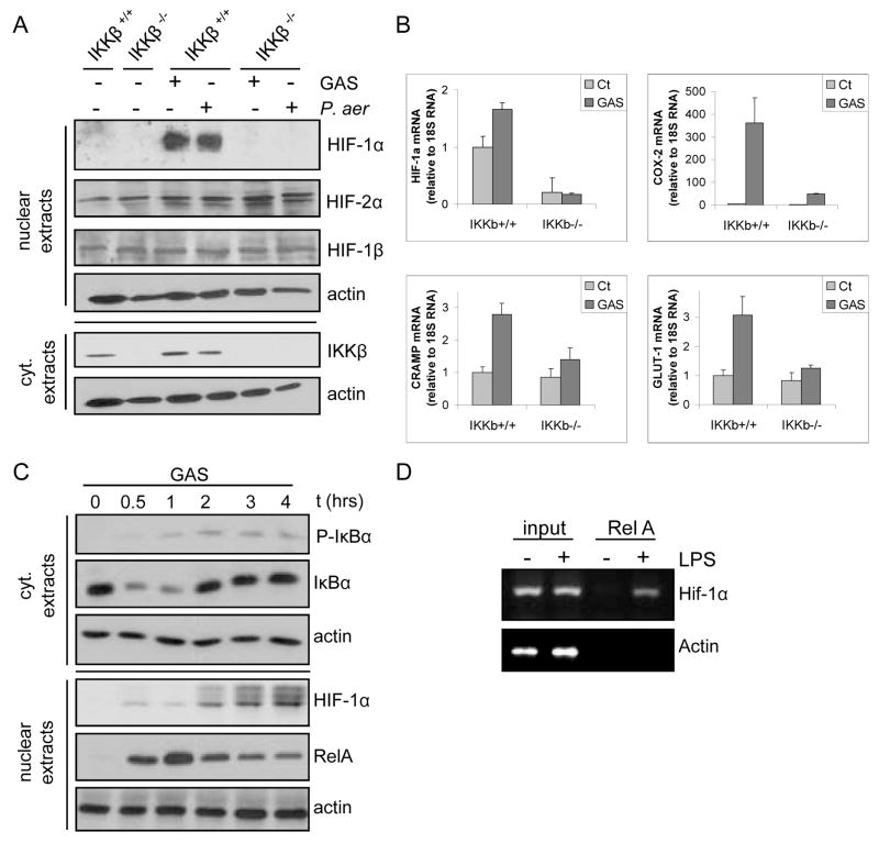
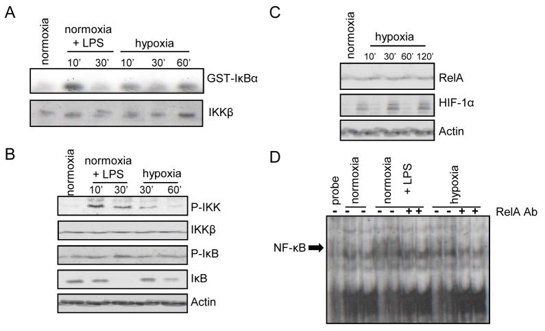
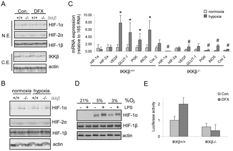
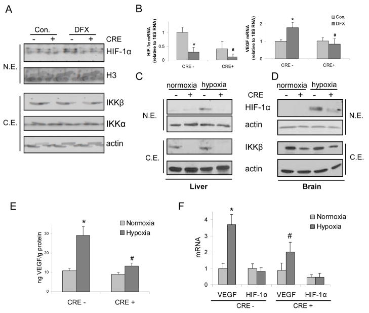
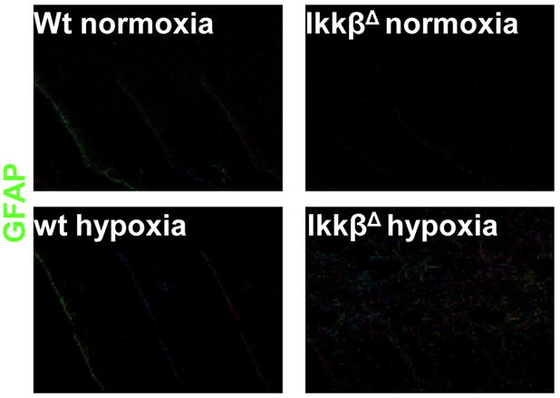
The hypoxic response is an ancient stress response triggered by low ambient oxygen (O2) (ref. 1) and controlled by hypoxia-inducible transcription factor-1 (HIF-1), whose alpha subunit is rapidly degraded under normoxia but stabilized when O2-dependent prolyl hydroxylases (PHDs) that target its O2-dependent degradation domain are inhibited. Thus, the amount of HIF-1alpha, which controls genes involved in energy metabolism and angiogenesis, is regulated post-translationally. Another ancient stress response is the innate immune response, regulated by several transcription factors, among which NF-kappaB plays a central role. NF-kappaB activation is controlled by IkappaB kinases (IKK), mainly IKK-beta, needed for phosphorylation-induced degradation of IkappaB inhibitors in response to infection and inflammation. IKK-beta is modestly activated in hypoxic cell cultures when PHDs that attenuate its activation are inhibited. However, defining the relationship between NF-kappaB and HIF-1alpha has proven elusive. Using in vitro systems, it was reported that HIF-1alpha activates NF-kappaB, that NF-kappaB controls HIF-1alpha transcription and that HIF-1alpha activation may be concurrent with inhibition of NF-kappaB. Here we show, with the use of mice lacking IKK-beta in different cell types, that NF-kappaB is a critical transcriptional activator of HIF-1alpha and that basal NF-kappaB activity is required for HIF-1alpha protein accumulation under hypoxia in cultured cells and in the liver and brain of hypoxic animals. IKK-beta deficiency results in defective induction of HIF-1alpha target genes including vascular endothelial growth factor. IKK-beta is also essential for HIF-1alpha accumulation in macrophages experiencing a bacterial infection. Hence, IKK-beta is an important physiological contributor to the hypoxic response, linking it to innate immunity and inflammation.


Figures

Figure 1
Figure 1. IKKβ is required for microbial-induced HIF-1α expression in macrophages
a) BMDM from either IkkβF/F (IKKβ+/+) or poly(IC)-injected IkkβF/F/Mx1-Cre (IkkβΔ; IKKβ−/−) mice were incubated with either with GAS or P. aeruginosa (MOI of 10 for 4 hrs). HIF-1α expression was analyzed by immunoblotting. b) RNA was extracted from BMDM incubated with GAS and gene expression was analyzed by quantitative (Q) RT-PCR. Results are averages of 3 separate experiments done in triplicate. Values were normalized relative to 18S rRNA. c) ChIP was performed with an anti-RelA antibody using fixed and sheared chromatin isolated from RAW264.7 mouse macrophages incubated with or without LPS. The HIF-1α promoter fragment, which contains a κB site at −197/−188 bp, was detected by PCR amplification.
Figure 2
Figure 2. Hypoxia activates the NF-kB pathway in macrophages
RAW264.7 mouse macrophages were incubated with or without LPS or cultured under hypoxia (O2 = 0.5 %). a) At the indicated time points of LPS stimulation or hypoxia, IKK activity was measured by an immunocomplex kinase assay using GST-IκBα as a substrate. b) Cell lysates were prepared and IKKβ and IκBα phosphorylation and abundance were analyzed by immunoblotting. c) Nuclear extracts were prepared at 2 hrs post-LPS or -hypoxia and NF-κB DNA binding activity was examined by a mobility shift assay. Antibody inhibition was performed using an anti-RelA antibody.
Figure 3
Figure 3. IKKβ regulates hypoxia-induced HIF-1α and target genes in mouse macrophages
a) BMDM from IkkβF/F (IKKβ+/+) or IkkβΔ (IKKβ−/−) mice were incubated with desferrioxamine (DFX) for 4 hrs. HIF-1α, HIF-1β and IKKβ expression were analyzed by immunoblotting. b) BMDM were obtained as above and cultured under hypoxia (O2 = 0.5% for 4 hrs). HIF-1α expression was analyzed by immunoblotting. c) BMDM were treated as above and mRNA expression was analyzed by Q-RT-PCR. Results are averages of three separate experiments done in triplicates. p<0.05: *, vs normoxic Ikkβ+/+ cells; #, vs hypoxic Ikkβ+/+ cells. d) MEF from either Ikkβ+/+ or Ikkβ−/− embryos were transfected with a luciferase reporter gene driven by the HIF-1α promoter. After 36 hrs the cells were incubated for 3 hrs with DFX. p<0.05: *, vs normoxic Ikkβ+/+ cells; #, vs hypoxic Ikkβ+/+ cells.
Figure 4
Figure 4. IKKβ regulates HIF-1α expression in hypoxic mice
IkkβF/F (CRE-) or IkkβΔ (CRE+) mice were treated with DFX (600 mg/Kg). After 15 hrs, livers were removed for protein (a) and RNA (b) analysis. a) HIF-1α and IKKβ expression was analyzed by immunoblotting. b) Expression of HIF-1α and VEGF mRNA was examined by Q-RT- PCR (n=3). p<0.05: *, vs normoxic CRE- mice; #, vs DFX-treated CRE- mice. c,d) IkkβF/F and IkkβΔ mice were kept under normoxia or hypoxia (O2 = 8%) for 24 hrs and HIF-1α expression was analyzed by immunoblotting of liver (c) or brain (d) extracts. e) VEGF expression in brain of mice from above experiment was analyzed by ELISA. p<0.05: *, vs normoxic CRE- mice; #, vs hypoxic CRE- mice. f) VEGF and HIF-1α mRNA expression was analyzed by Q-RT-PCR of total brain RNA. p<0.05: *, vs normoxic CRE- mice; #, vs hypoxic CRE- mice (n=3).
Figure 5
Figure 5. IKKβ deficiency results in increased astrogliosis in brains of hypoxic mice
Mice of the indicated genotypes were kept under normoxia or hypoxia (O2 = 8%) for 24 hrs. After this period the mice were perfused with a fixative and the brain was collected and frozen. Brain sections at the cerebellar region (10 μm) were stained with an antibody against GFAP (an astrocyte marker). Magnification x20.

References

    1. Wang GL, Semenza GL. General involvement of hypoxia-inducible factor 1 in transcriptional response to hypoxia. Proc Natl Acad Sci U S A. 1993;90:4304–4308. - PMC - PubMed
    1. Maxwell PH, MSW, GWC, SCC, ECV, MEC, Wykoff CC, Pugh CW, Maher ER, Ratcliffe PJ. The tumour suppressor protein VHL targets hypoxia-inducible factors for oxygen-dependent proteolysis. Nature. 1999;399:271–275. - PubMed
    1. Semenza G. HIF-1, O(2), and the 3 PHDs: how animal cells signal hypoxia to the nucleu. Cell. 2001;107:1–3. - PubMed
    1. Schofield CJ, Ratcliffe PJ. Oxygen sensing by HIF hydroxylase. Nat Rev Mol Cell Biol. 2004;5:343–354. - PubMed
    1. Akira S, Uematsu S, Takeuchi O. Pathogen recognition and innate immunity. Cell. 2006;124:783–801. - PubMed

Publication types

MeSH terms