. 2008 May 1;453(7191):102-5.
doi: 10.1038/nature06829. Epub 2008 Apr 23.

Melanopsin cells are the principal conduits for rod-cone input to non-image-forming vision

Affiliations

Melanopsin cells are the principal conduits for rod-cone input to non-image-forming vision

Ali D Güler et al. Nature. .

Abstract

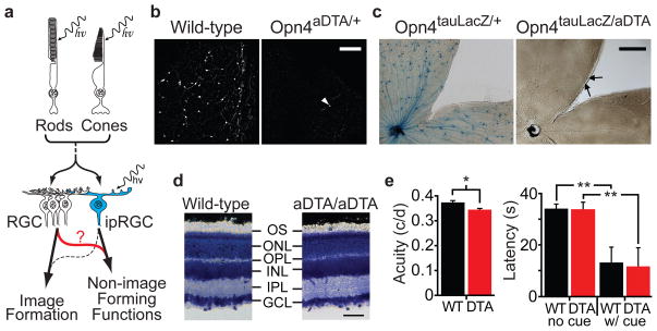
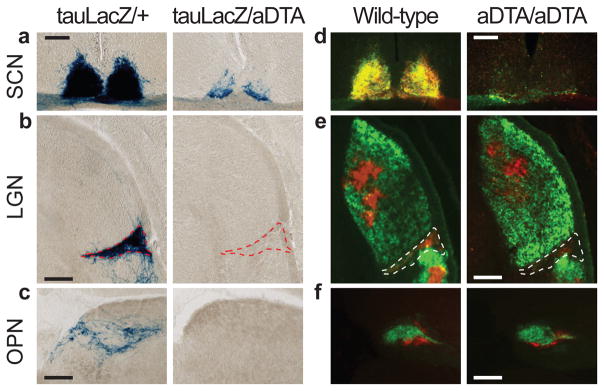
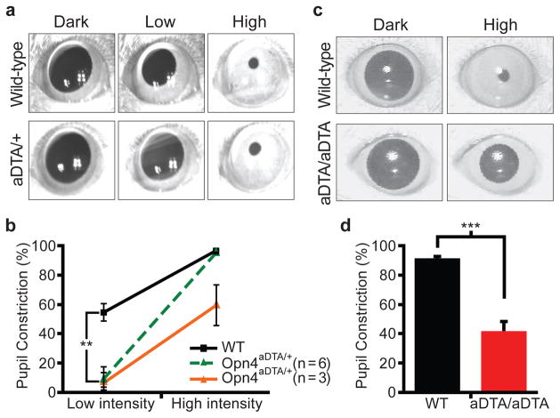
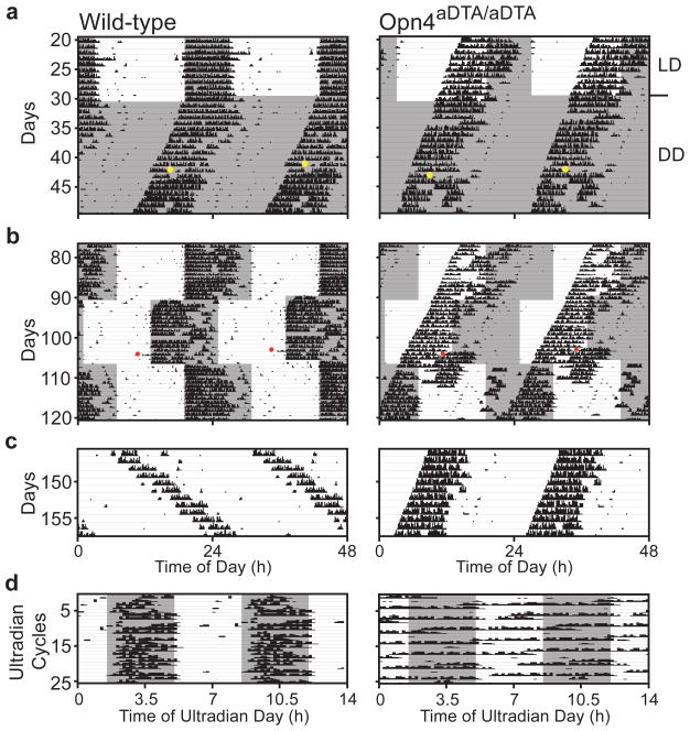
Rod and cone photoreceptors detect light and relay this information through a multisynaptic pathway to the brain by means of retinal ganglion cells (RGCs). These retinal outputs support not only pattern vision but also non-image-forming (NIF) functions, which include circadian photoentrainment and pupillary light reflex (PLR). In mammals, NIF functions are mediated by rods, cones and the melanopsin-containing intrinsically photosensitive retinal ganglion cells (ipRGCs). Rod-cone photoreceptors and ipRGCs are complementary in signalling light intensity for NIF functions. The ipRGCs, in addition to being directly photosensitive, also receive synaptic input from rod-cone networks. To determine how the ipRGCs relay rod-cone light information for both image-forming and non-image-forming functions, we genetically ablated ipRGCs in mice. Here we show that animals lacking ipRGCs retain pattern vision but have deficits in both PLR and circadian photoentrainment that are more extensive than those observed in melanopsin knockouts. The defects in PLR and photoentrainment resemble those observed in animals that lack phototransduction in all three photoreceptor classes. These results indicate that light signals for irradiance detection are dissociated from pattern vision at the retinal ganglion cell level, and animals that cannot detect light for NIF functions are still capable of image formation.


Figures

Figure 1
Figure 1. Elimination of ipRGCs in mouse retina
a, Model describing how rod/cone signalling through conventional RGCs or ipRGCs contribute to NIF functions. Role of ipRGCs in image formation is speculative (dotted line). b, Melanopsin antibody staining in retinas of 18 month old wild-type (n=6) and Opn4aDTA/+ (n=12) mice. White arrowhead indicates a surviving ipRGC. Scale bar, 200μm. c, X-gal staining from Opn4tau-LacZ/+ (n=6) and Opn4aDTA/tau-LacZ (n=8). The surviving cells are weakly stained (black arrows). Scale bar, 500μm. d, Cross sections of Giemsa stained retinas from 18 month old Opn4aDTA/aDTA (aDTA/aDTA; n=3) and wild-type mice (n=3). The morphology of retinas is indistinguishable between genotypes. GCL, ganglion cell layer; IPL, inner plexiform layer; INL, inner nuclear layer; OPL, outer plexiform layer; ONL, outer nuclear layer; OS, outer segment. Scale bar, 50 μm. e, Left, the acuity of Opn4aDTA/aDTA mice (DTA; n=11; red bar) was slightly decreased compared to wild types (WT; n=9; black bar). Right, the latency to locate a marked platform (w/cue) in a water maze was similar between Opn4aDTA/aDTA (DTA; n=14; red bar) and wild types (WT; n=12; black bar). This latency significantly differed for unmarked platform (no cue) tests. All statistical comparisons utilized Student’s t test (*, p<0.05; **, p<0.01); error bars ± s.e.m.
Figure 2
Figure 2. The ipRGC fibres in the brain decrease in aDTA mice
a, b, and c, X-gal staining in Opn4tau-LacZ/aDTA (tauLacZ/aDTA; n=2) show that ipRGC innervation of the SCN, IGL and OPN is decreased. d, e, and f, Ocular cholera toxin injections (left eye, green; right, red) of Opn4aDTA/aDTA (aDTA/aDTA; n=11) and wild types (n=6). a and d, SCN innervation is sparse in aDTA mice. b and e, The dorsal lateral geniculate nucleus (LGN) is innervated similarly both in aDTA and wild-type animals, while few fibres remain in the IGL of mutant mice (outlined region). c and f, The OPN shell is innervated by ipRGCs and the core is targeted by other RGCs. f, Fibres in the OPN core are retained in Opn4aDTA/aDTA mice. c, Fibres in the shell region are eliminated in Opn4tau-LacZ/aDTA animals. Scale bars, 200μm.
Figure 3
Figure 3. Opn4aDTA mice have deficits in PLR
a, All 9 Opn4aDTA/+ mice showed defective PLR (1.8μW/cm2; 30s white light; Low) that induces 50% constriction in wild types. The Opn4aDTA/+ mice showed a 9.0±6.0% constriction. 6 of 9 Opn4aDTA/+ mice had full pupil constriction at high light intensity (3mW/cm2; High). The rest of the mutant mice (3 of 9) showed defective PLR. b, Quantification of PLR data of wild-type (WT; n=9; black squares) and Opn4aDTA/+ either photoentrained (green triangles; n=6) or non-photoentrained (orange triangles; n=3) animals. All statistical comparisons were made by Student’s t test (**, p<0.01). c, All Opn4aDTA/aDTA animals constrict their pupil only to a maximum of 42% at a light pulse that causes 95% constriction in wild types (161μW/cm2, 470nm monochromatic light; High). d, Quantification of PLR data of wild-type (WT; n=11; black bar) and Opn4aDTA/aDTA (aDTA/aDTA; n=12; red bar) mice from c. Statistical comparisons were made by Student’s t test (***, p<0.001); error bars ± s.e.m.
Figure 4
Figure 4. Opn4aDTA/aDTA mice do not photoentrain or mask
a, Opn4aDTA/aDTA mice free-run under light/dark cycles (grey and white backgrounds; dark and light (~700lux), respectively). Opn4aDTA/aDTA mice do not phase shift to a 15-minute 1500lux white light pulse (CT16; yellow dots: light pulses). b, Opn4aDTA/aDTA mice do not photoentrain to the 24-hour light/dark cycle in the delay or advance phases. c, Unlike wild-type animals, no Opn4aDTA/aDTA mice lengthened their period under constant light. d, Opn4aDTA/aDTA mice do not mask under 7-hour ultradian cycle. Red dots: cage changes.

References

    1. Hubel DH. Eye, brain, and vision (Scientific American Library) W.H. Freeman; New York: 1988.
    1. Berson DM, Dunn FA, Takao M. Phototransduction by retinal ganglion cells that set the circadian clock. Science. 2002;295:1070–3. - PubMed
    1. Hattar S, Liao HW, Takao M, Berson DM, Yau KW. Melanopsin-containing retinal ganglion cells: architecture, projections, and intrinsic photosensitivity. Science. 2002;295:1065–70. - PMC - PubMed
    1. Freedman MS, et al. Regulation of mammalian circadian behavior by non-rod, non-cone, ocular photoreceptors. Science. 1999;284:502–4. - PubMed
    1. Czeisler CA, et al. Suppression of melatonin secretion in some blind patients by exposure to bright light. N Engl J Med. 1995;332:6–11. - PubMed

MeSH terms