. 2008 Apr 21:14:675-82.

A novel splice-site mutation of TULP1 underlies severe early-onset retinitis pigmentosa in a consanguineous Israeli Muslim Arab family

Affiliations

A novel splice-site mutation of TULP1 underlies severe early-onset retinitis pigmentosa in a consanguineous Israeli Muslim Arab family

Anan H Abbasi et al. Mol Vis. .

Abstract

Purpose: To investigate the genetic basis for autosomal recessive severe early-onset retinitis pigmentosa (RP) in a consanguineous Israeli Muslim Arab family.

Methods: Haplotype analysis for all known genes underlying autosomal recessive RP was performed. Mutation screening of the underlying gene was done by direct sequencing. An in vitro splicing assay was used to evaluate the effect of the identified mutation on splicing.

Results: Haplotype analysis indicated linkage to the Tubby-like protein 1 (TULP)1 gene. Direct sequencing revealed a homozygous single base insertion, c.1495+2_1495+3insT, located in the conserved donor splice-site of intron 14. This mutation co-segregated with the disease, and was not detected in 114 unrelated Israeli Muslim Arab controls. We used an in vitro splicing assay to demonstrate that this mutation leads to incorrect splicing.

Conclusions: To date, 22 distinct pathogenic mutations of TULP1 have been reported in patients with early-onset RP or Leber congenital amaurosis. Here we report a novel splice-site mutation of TULP1, c.1495+2_1495+3insT, underlying autosomal recessive early-onset RP in a consanguineous Israeli Muslim Arab family. This report expands the spectrum of pathogenic mutations of the TULP1 gene.


Figures

Figure 1
Figure 1
Pedigree and haplotype analysis. Shown is a pedigree of a consanguineous Israeli Muslim Arab family segregating early-onset autosomal recessive retinitis pigmentosa (family TB13). Haplotype analysis performed on this family demonstrated co-segregation of a haplotype of three polymorphic marker alleles, linked to TULP1 on chromosome 6p21.31, with autosomal recessive retinitis pigmentosa in this family. The mutation-bearing haplotype is marked by a box. Males are represented by squares, females are represented by circles. Affected individuals are marked by black shadings.
Figure 2
Figure 2
Fundus photographs of individual IV-18 (right eye). The photographs show vascular attenuation, peripheral bone spicule pigmentation, accompanied by salt and pepper-like changes, and signs of maculopathy, including edema and a yellow perifoveal ring.
Figure 3
Figure 3
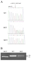
The c.1495+2_1495+3insT mutation. A: Shown are nucleotide sequence traces of the boundary between TULP1 exon and intron14 in a non-carrier individual (wt), an individual heterozygote for the c.1495+2_1495+3insT mutation (het), and an affected individual homozygote for the mutant allele (mut). The exon-intron boundary is marked. B: A mismatch-primer restriction–based assay used to screen control DNA samples for the c.1495+2_1495+3insT mutation. The reverse PCR primer included one mismatched base, which, in combination with the mutant allele, but not with the WT allele, created a DrdI restriction site. A 286 bp PCR product derived from the WT allele was not digested by DrdI, while in the presence of the MUT two restriction fragments of 251 and 35 bp were obtained. Digestion of a PCR product derived from a heterozygote for the mutation (HET) yielded three fragments (286, 251, and 35 bp). The 35 bp fragment is not visible.
Figure 4
Figure 4
Minigene constructs and products obtained in the in vitro splicing assay. A: Shown is a schematic representation of the constructs, which include TULP1 exons 12 to 15 (represented by boxes), flanked by 79–183 bp of intronic sequences (represented by straight lines). Constructs harbored either the wild-type (WT) or one of two mutant alleles at the donor splice-site of intron 14: c.1495+2_1495+3insT and c.1495+1G>A. Also shown are the locations of primers used for RT–PCR analysis (indicated by arrows) and the obtained splicing products. Sizes of exonic and intronic fragments are indicated below them. B: WT and mutant constructs were transfected into Y79 cells, followed by RNA extraction and RT–PCR analysis. No PCR products were obtained from cells transfected with the empty pCMV-script vector. β-actin (ACTB), a 437 bp product, served as an internal control for RNA quality and quantity.

References

    1. Fazzi E, Signorini SG, Scelsa B, Bova SM, Lanzi G. Leber's congenital amaurosis: an update. Eur J Paediatr Neurol. 2003;7:13–22. - PubMed
    1. Hanein S, Perrault I, Gerber S, Tanguy G, Barbet F, Ducroq D, Calvas P, Dollfus H, Hamel C, Lopponen T, Munier F, Santos L, Shalev S, Zafeiriou D, Dufier JL, Munnich A, Rozet JM, Kaplan J. Leber congenital amaurosis: comprehensive survey of the genetic heterogeneity, refinement of the clinical definition, and genotype-phenotype correlations as a strategy for molecular diagnosis. Hum Mutat. 2004;23:306–17. - PubMed
    1. Koenekoop RK. An overview of Leber congenital amaurosis: a model to understand human retinal development. Surv Ophthalmol. 2004;49:379–98. - PubMed
    1. Hartong DT, Berson EL, Dryja TP. Retinitis pigmentosa. Lancet. 2006;368:1795–809. - PubMed
    1. Rivolta C, Sharon D, DeAngelis MM, Dryja TP. Retinitis pigmentosa and allied diseases: numerous diseases, genes, and inheritance patterns. Hum Mol Genet. 2002;11:1219–27. - PubMed

Publication types

MeSH terms

LinkOut - more resources