. 2008 Apr 24:8:45.
doi: 10.1186/1471-213X-8-45.

Rex1/Zfp42 is dispensable for pluripotency in mouse ES cells

Affiliations

Rex1/Zfp42 is dispensable for pluripotency in mouse ES cells

Shinji Masui et al. BMC Dev Biol. .

Abstract

Background: Rex1/Zfp42 has been extensively used as a marker for the undifferentiated state of pluripotent stem cells. However, its function in pluripotent stem cells including embryonic stem (ES) cells remained unclear although its involvement in visceral endoderm differentiation in F9 embryonal carcinoma (EC) cells was reported.

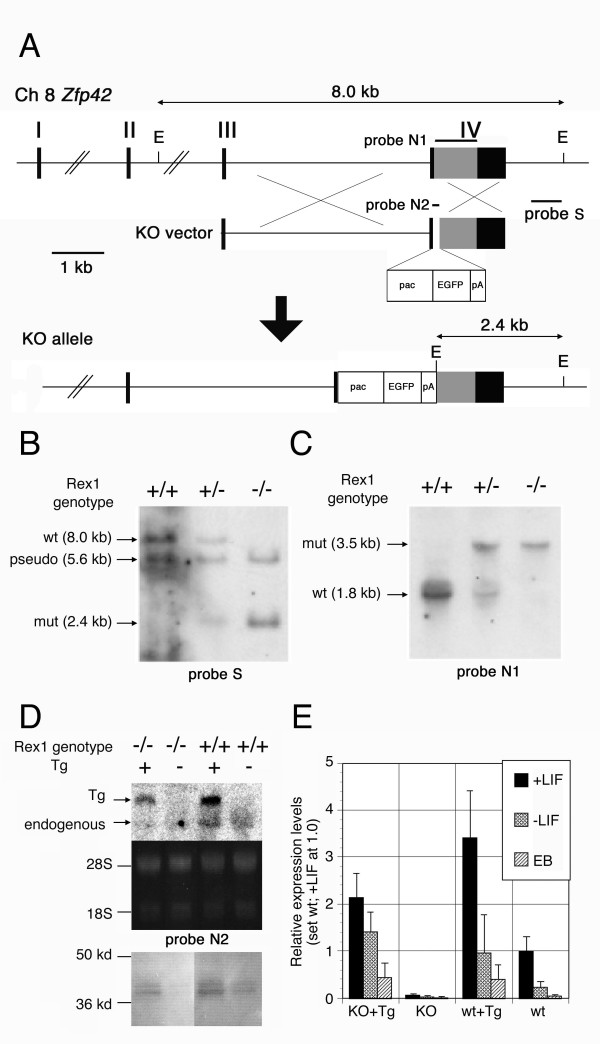
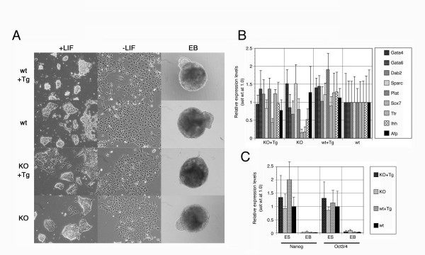
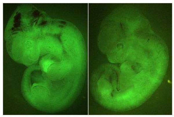
Results: We showed the function of Rex1 in mouse ES cells as well as in embryos using the conventional gene targeting strategy. Our results clearly indicated that Rex1 function is dispensable for both the maintenance of pluripotency in ES cells and the development of embryos. However, Rex1-/- ES cells showed the defect to induce a subset of the marker genes of visceral endoderm, when differentiated as embryoid body, as found in EC cells.

Conclusion: Rex1 should be regarded just as a marker of pluripotency without functional significance like the activity of alkaline phosphatase.


Figures

Figure 1
Figure 1
Generation of ES cells with various Rex1 genotypes. A. Strategy for generation of Rex1-KO ES cells. The schematic maps of the Rex1 allele (top), the KO vector carrying the floxed pacEGFP-pA cassette (middle), and the KO allele generated by homologous recombination (bottom) were shown in scale. The EcoRI sites (E) provide the polymorphism between the wild-type and mutant alleles, 8.0 kb and 2.4 kb, respectively, on southern blot analysis using the indicated 3' external probe. B. Southern blot hybridization of wild-type (+/+), Rex1 heterozygous (+/-) and homozygous (-/-) ES cells using the EcoRI digestion and the 3' external probe. The expected sizes of wild-type (wt) and mutant (mut) bands were detected. The 5.6 kb fragment corresponds to the polymorphism of the Rex1 pseudogene on chromosome 15 reported previously as well as found in the mouse genome data. C. Northern blot analysis of Rex1 expression in wild-type (+/+), Rex1 heterozygous (+/-) and homozygous (-/-) ES cells. The Rex1 cDNA probe detects 1.8 kb mRNA from the wild-type allele and 3.5 kb mRNA, which is generated by inefficient function of the polyA addition signal in the pacEGFP-pA cassette, from the mutant allele. The Rex1 KO ES cells lack the wild-type transcript. D. Northern and western blot analysis of wild-type (+/+) and Rex1 KO (-/-) ES cells with the Rex1 transgene (Tg:+) or the empty vector (Tg:-). The 2.7 kb transcripts from the transgene were detected with or without the 2.2 kb endogenous transcripts in Northern blot with the Rex1 cDNA probe (top), in which equal loading of total RNA was confirmed by ethidium bromide staining of 28S and 18S ribosomal RNAs (middle). Western blot using anti-Rex1 antisera detects ~38 kd band in wild-type, wild-type+Tg and Rex1 KO+Tg lanes but not Rex1 KO lane (bottom), confirming the proper production of Rex1 protein from Tg. E. QPCR analysis of Rex1 expression in undifferentiated (+LIF) and differentiated (-LIF and EB) ES cells with various Rex1 genotypes. Three independent clones with each genotypes were cultured with or without LIF for 4 days or for formation of EBs for 5 days, analyzed separately with normalization by the amount of Gapdh, and plotted with standard deviation against the expression level in undifferentiated wild-type ES cells (wt) cultured with LIF, set as 1.0. The primer pair was set in the region deleted in the KO allele.
Figure 2
Figure 2
Nuclear localization of Rex1. The expression vectors of EGFP-tagged or HA-tagged Rex1 were transiently transfected into HeLa and ES cells, and the localization of these transgene products were detected by fluorescent microscopy directly (for EGFPRex1) or after immunostaining for HA-tag (for HARex1). Phase contrast (left), fluorescent (middle) and their merged image (right) were shown for each transfectants. The fluorescent signals were localized in nuclei in both HeLa and ES cells for both chimeric Rex1 proteins.
Figure 3
Figure 3
Gene expression profile in Rex1 KO ES cells. A. DNA microarray analysis of Rex1 KO ES cells. Scatter-plot of log-ratios of relative expression levels were shown for wild-type (EB5) versus Rex1 KO (HP3) ES cells. B. QPCR analyses of expressions of the putative Rex1 target genes. Three independent clones with each genotypes were cultured with LIF for 4 days, analyzed separately with normalization by the amount of Gapdh, and plotted with standard deviation against the expression level in undifferentiated wild-type ES cells (wt), set as 1.0.
Figure 4
Figure 4
Differentiation of ES cells with various Rex1 genotypes. A. Photomicrographs of colonies at 4 days cultured with LIF (left) or without LIF (middle), or EBs at 5 days (right) derived from representative ES cells with indicated genotypes for Rex1. Scale bar is 100 μm. B. QPCR analyses of the endoderm marker genes in EBs derived from ES cells with various Rex1 genotypes. Three independent clones with each genotypes were cultured for formation of EBs for 5 days, analyzed separately with normalization by the amount of Gapdh, and plotted with standard deviation against the expression level in wild-type ES cells-derived EBs, set as 1.0. C. QPCR analyses of the stem cell marker genes in ES cells and EBs with various Rex1 genotypes. Three independent clones with each genotypes were cultured for 4 days with LIF or for formation of EBs for 5 days, analyzed separately with normalization by the amount of Gapdh, and plotted with standard deviation against the expression level in wild-type ES cells, set as 1.0.
Figure 5
Figure 5
Chimeric embryos derived from Rex1 KO ES cells. When HP4-EGFP ES cells, which were homozygotes for the mutant Rex1 allele and marked by the constitutively-active Egfp transgene, were injected into blastocysts, the embryos developed to chimeras at 12.5 dpc in which widespread contributions of GFP-positive cells were observed in fluorescent microscopic observation.

Similar articles

Cited by

References

    1. Boyer LA, Mathur D, Jaenisch R. Molecular control of pluripotency. Curr Opin Genet Dev. 2006;16:455–62. doi: 10.1016/j.gde.2006.08.009. - DOI - PubMed
    1. Niwa H. How is pluripotency determined and maintained? Development. 2007;134:635–46. doi: 10.1242/dev.02787. - DOI - PubMed
    1. Nichols J, Zevnik B, Anastassiadis K, Niwa H, Klewe-Nebenius D, Chambers I, Scholer H, Smith A. Formation of pluripotent stem cells in the mammalian embryo depends on the POU transcription factor Oct4. Cell. 1998;95:379–91. doi: 10.1016/S0092-8674(00)81769-9. - DOI - PubMed
    1. Niwa H, Miyazaki J, Smith AG. Quantitative expression of Oct-3/4 defines differentiation, dedifferentiation or self-renewal of ES cells. Nat Genet. 2000;24:372–6. doi: 10.1038/74199. - DOI - PubMed
    1. Avilion AA, Nicolis SK, Pevny LH, Perez L, Vivian N, Lovell-Badge R. Multipotent cell lineages in early mouse development depend on SOX2 function. Genes Dev. 2003;17:126–40. doi: 10.1101/gad.224503. - DOI - PMC - PubMed

Publication types

MeSH terms