. 2008 Apr 24:6:10.
doi: 10.1186/1741-7015-6-10.

Clinical characterization and the mutation spectrum in Swedish adenomatous polyposis families

Affiliations

Clinical characterization and the mutation spectrum in Swedish adenomatous polyposis families

Gunilla Kanter-Smoler et al. BMC Med. .

Abstract

Background: The dominantly inherited condition familial adenomatous polyposis (FAP) is caused by germline mutations in the APC gene. Finding the causative mutations has great implications for the families. Correlating the genotypes to the phenotypes could help to improve the diagnosis and follow-up of patients.

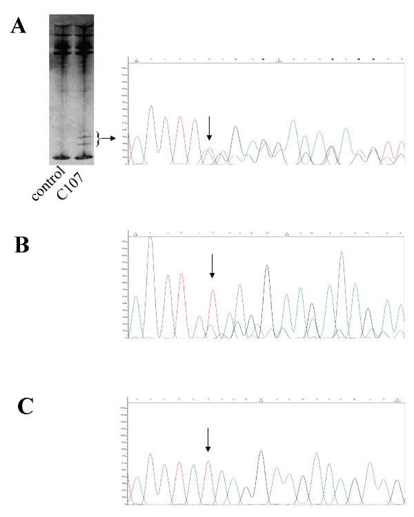
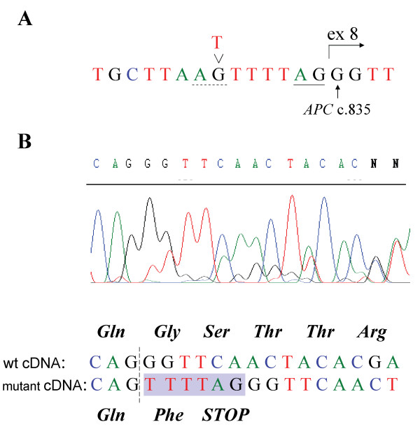
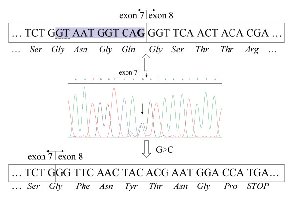
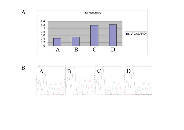
Methods: Mutation screening of APC and the clinical characterization of 96 unrelated FAP patients from the Swedish Polyposis Registry was performed. In addition to generally used mutation screening methods, analyses of splicing-affecting mutations and investigations of the presence of low-frequency mutation alleles, indicating mosaics, have been performed, as well as quantitative real-time polymerase chain reaction to detect lowered expression of APC.

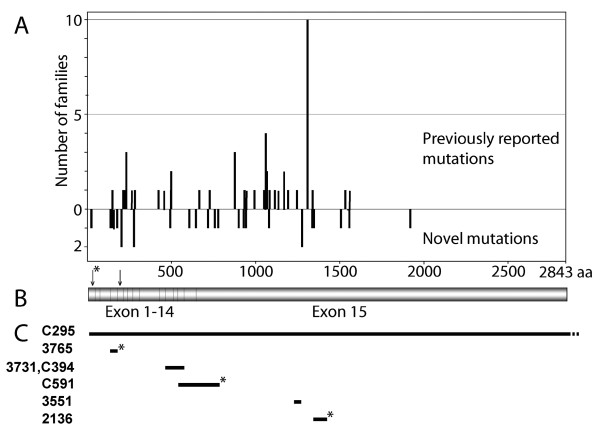
Results: Sixty-one different APC mutations in 81 of the 96 families were identified and 27 of those are novel. We have previously shown that 6 of the 96 patients carried biallelic MUTYH mutations. The 9 mutation-negative cases all display an attenuated or atypical phenotype. Probands with a genotype (codon 1250-1464) predicting a severe phenotype had a median age at diagnosis of 21.8 (range, 11-49) years compared with 34.4 (range, 14-57) years among those with mutations outside this region (P < 0.017). Dense polyposis (> 1000) occurred in 75% of the probands with a severe phenotype compared with 30% in those with mutations outside this region. The morbidity in colorectal cancer among probands was 25% at a mean age of 37.5 years and 29% at a mean age of 46.6 years.

Conclusion: Using a variety of mutation-detection techniques, we have achieved a 100% detection frequency in classical FAP. Probands with APC mutations outside codon 1250-1464, although exhibiting a less-severe phenotype, are at high risk of having a colorectal cancer at diagnosis indicating that age at diagnosis is as important as the severity of the disease for colorectal cancer morbidity.


Figures

Figure 1
Figure 1
Mutation spectrum of the APC gene. (A) The spectrum of APC mutations identified among families from the Swedish Polyposis Registry showing the distribution between previously reported and novel mutations in our patients. (B) A schematic representation of the APC coding region, shown in the same scale as in (A). The arrow with an asterisk indicates codon 24 and the second arrow points at codon 184. (C) Distribution of six large deletions found in seven unrelated patients of the Swedish Polyposis Registry. Novel deletions are marked with an asterisk. Patient numbers are shown to the left. Scale as in (A).
Figure 2
Figure 2
Detection of the mosaic c.2700_2701delTC mutation in patient C107. Nucleotide 2700 is indicated with an arrow. (A) The aberrant bands indicated by the bracket were excised from the SSCP/HD gel. The resulting DNA sequence is shown to the right. (B) DNA sequence from DNA extracted from tumor-derived cells from the patient. (C) The DNA sequence from DNA isolated from the patient's blood lymphocytes.
Figure 3
Figure 3
Characterization of the mutation in patient C496. (A) Genomic sequence of the patient showing the c.835-7T > G mutation. The new splice site generated by the T > G substitution is indicated with a dashed line, the wildtype acceptor-splice site is underlined, and the regular start of exon 8 is indicated with an arrow. (B) cDNA sequence covering the exon 7–8 boundary, indicated with a dashed line. Shown below the sequence diagram is the interpretation of the sequence reflecting the two mRNA species present in the sample. The insertion of 6 bp owing to the introduction of a new splice site in the mutant allele is shown as a shaded area. Predicted amino-acid sequence of translation products are shown above and below the respective cDNA sequence.
Figure 4
Figure 4
Characterization of mutation in patient C633. Diagram of genomic DNA sequence at the exon/intron 7 boundary. The line arrow indicates the c.834G > C mutation and the wildtype 5' donor splice site of intron 7 is underlined in the sequence diagram. The wildtype cDNA and the resulting amino acid sequence from the corresponding transcript are shown above the diagram. The G that is substituted in one allele in the patient is indicated in bold. The cryptic splice site used as a result of the mutation is underlined with a dashed line and the shaded area corresponds to the mRNA sequence deleted in the mutant transcript. Beneath the genomic sequence the cDNA sequence derived from the mutant allele is displayed, showing the resulting frameshift and premature termination of the translation product.
Figure 5
Figure 5
mRNA expression analysis of family 1 of the Swedish Polyposis Registry. (A) Diagram of part of the results from the TaqMan APC mRNA expression analysis, showing the relative mRNA levels calculated by the standard curve method of two affected members of family 1 (A and B) and two control individuals (C and D). (B) Diagrams of cDNA sequences of the above indicated patients and controls covering the APC c.5465A > T polymorphism.
Figure 6
Figure 6
Pedigree presenting a part of family 1 of the Swedish Polyposis Registry. Family members where positive linkage to APC has been confirmed are indicated with asterisks.

References

    1. Groden J, Thliveris A, Samowitz W, Carlson M, Gelbert L, Albertsen H, Joslyn G, Stevens J, Spirio L, Robertson M, Sargeant L, Krapcho K, Wolff BurtdER, Hughes JP, Warrington J, McPherson J, Wasmuth J, Le Paslier D, Abderrahim H, Cohen C, Leppert M, White R. Identification and characterization of the familial adenomatous polyposis coli gene. Cell. 1991;66:589–600. doi: 10.1016/0092-8674(81)90021-0. - DOI - PubMed
    1. Kinzler KW, Nilbert MC, Vogelstein B, Bryan TM, Levy DB, Smith KJ, Preisinger AC, Hamilton SR, Hedge P, Markham A, Carlson M, Joslyn G, Groden J, White R, Miki Y, Miyoshi Y, Nishisho I, Nakamura Y. Identification of a gene located at chromosome 5q21 that is mutated in colorectal cancers. Science. 1991;251:1366–1370. doi: 10.1126/science.1848370. - DOI - PubMed
    1. Lipton L, Halford SE, Johnson V, Novelli MR, Jones A, Cummings C, Barclay E, Sieber O, Sadat A, Bisgaard ML, Hodgson SV, Aaltonen LA, Thomas HJ, Tomlinson IP. Carcinogenesis in MYH-associated polyposis follows a distinct genetic pathway. Cancer Res. 2003;63:7595–7599. - PubMed
    1. Al-Tassan N, Chmiel NH, Maynard J, Fleming N, Livingston AL, Williams GT, Hodges AK, Davies DR, David SS, Sampson JR, Cheadle JP. Inherited variants of MYH associated with somatic G:C-->T:A mutations in colorectal tumors. Nat Genet. 2002;30:227–232. doi: 10.1038/ng828. - DOI - PubMed
    1. Jones S, Emmerson P, Maynard J, Best JM, Jordan S, Williams GT, Sampson JR, Cheadle JP. Biallelic germline mutations in MYH predispose to multiple colorectal adenoma and somatic G:C-->T:A mutations. Hum Mol Genet. 2002;11:2961–2967. doi: 10.1093/hmg/11.23.2961. - DOI - PubMed

Publication types