. 2008 Jun;135(11):1969-79.
doi: 10.1242/dev.021444. Epub 2008 Apr 23.

PAP- and GLD-2-type poly(A) polymerases are required sequentially in cytoplasmic polyadenylation and oogenesis in Drosophila

Affiliations

PAP- and GLD-2-type poly(A) polymerases are required sequentially in cytoplasmic polyadenylation and oogenesis in Drosophila

Perrine Benoit et al. Development. 2008 Jun.

Abstract

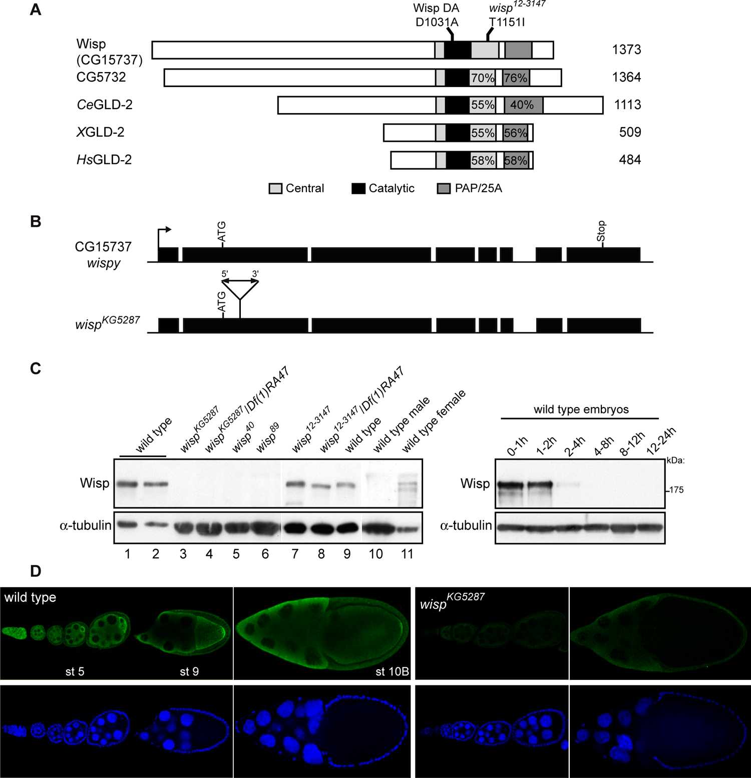
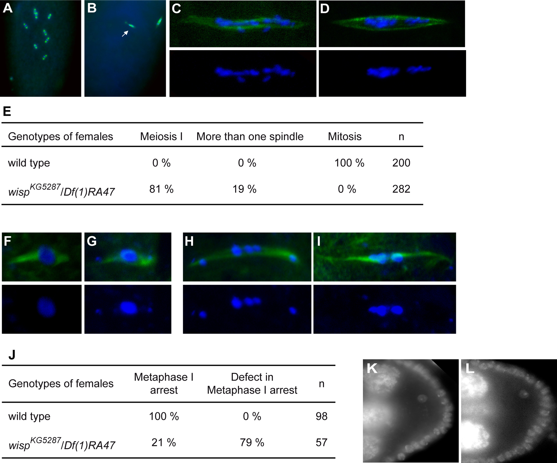
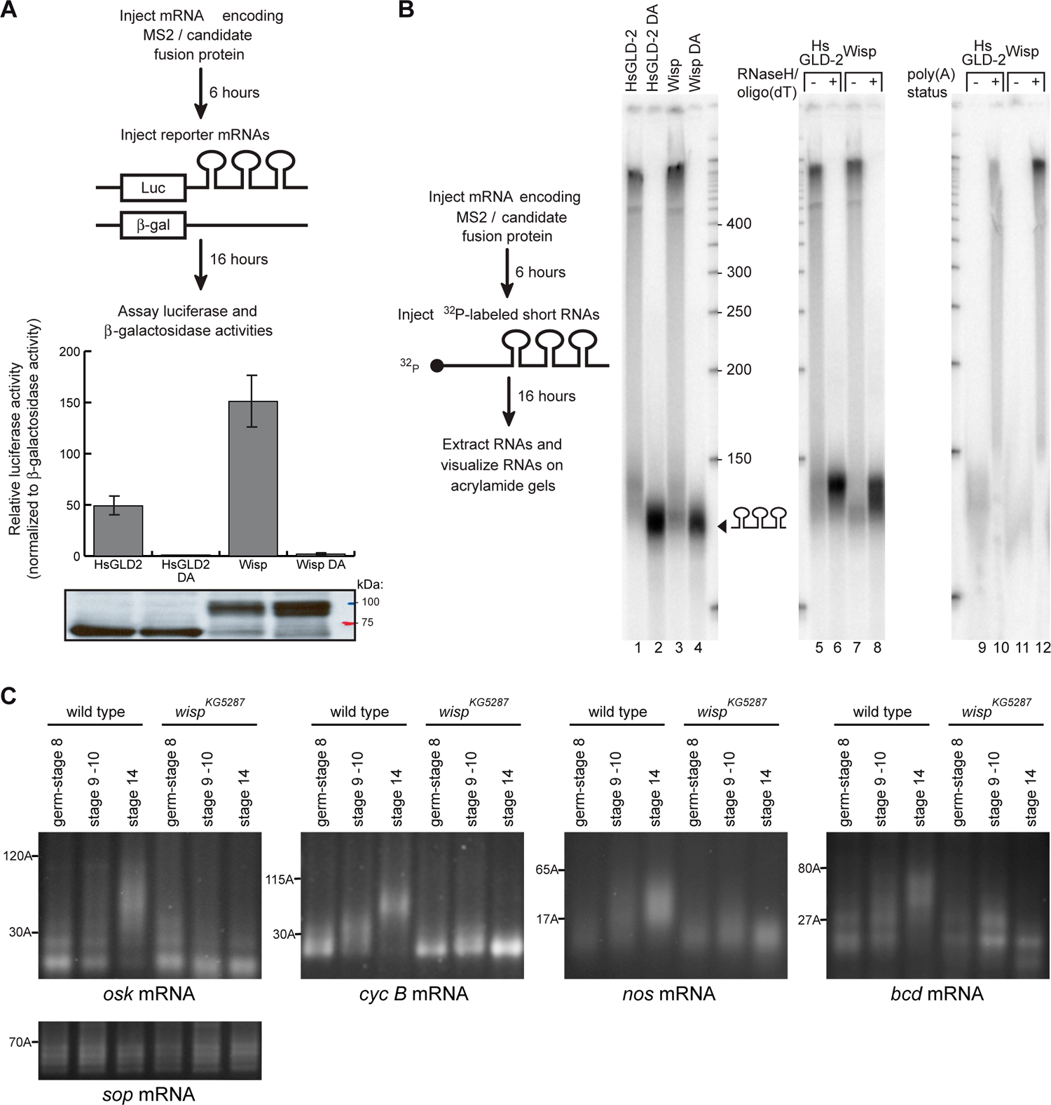
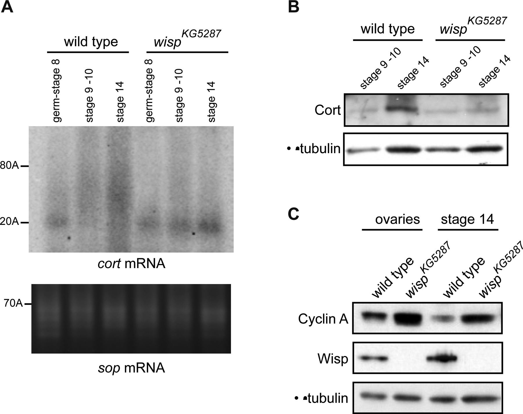
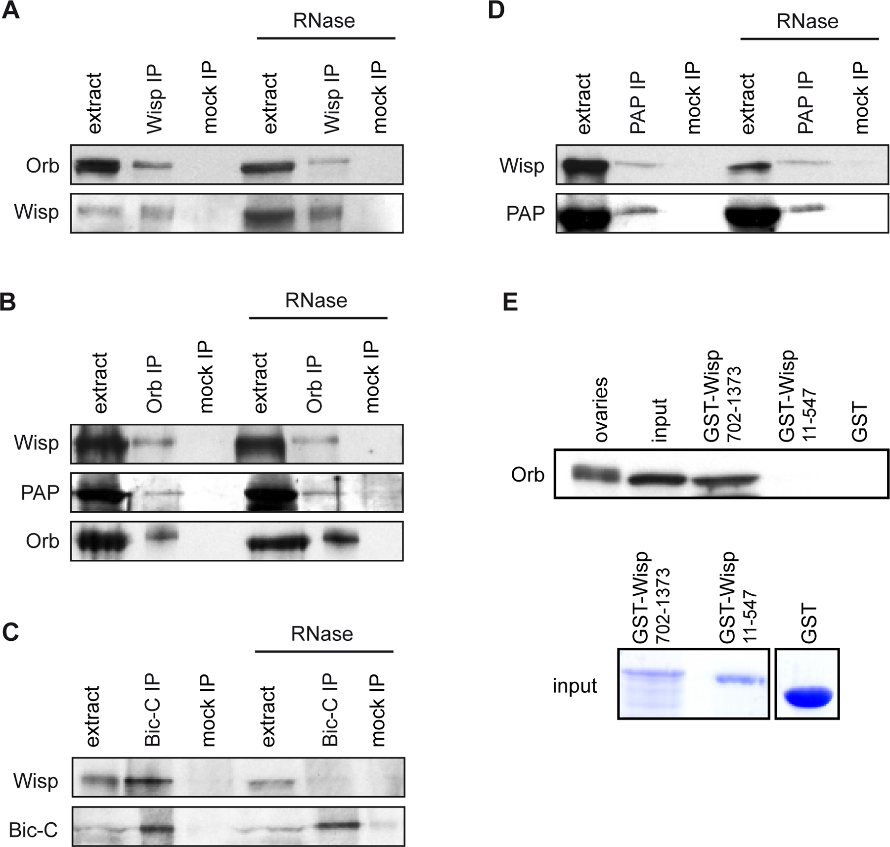
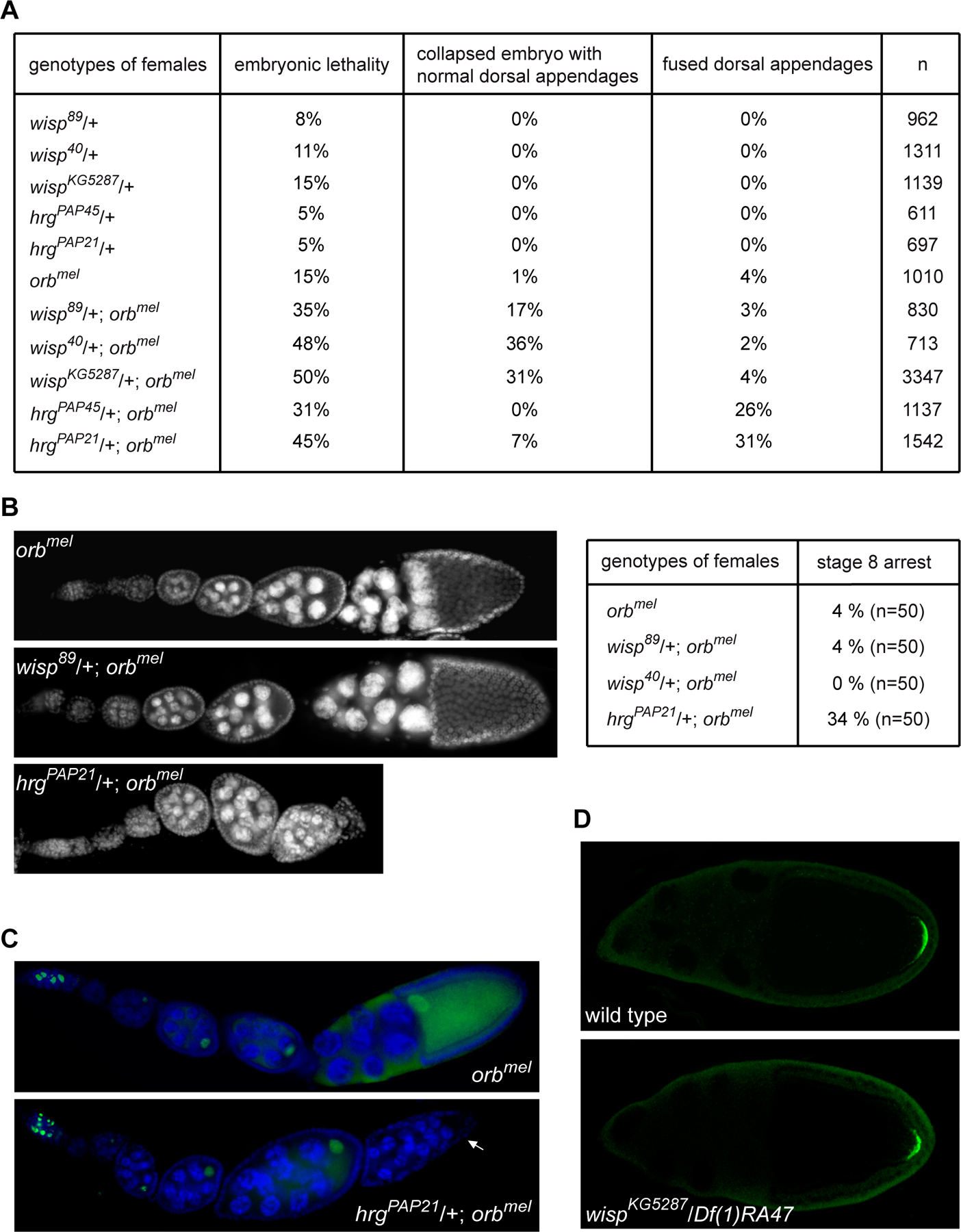
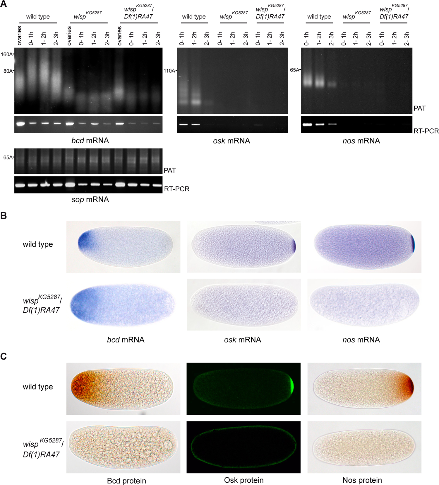
Cytoplasmic polyadenylation has an essential role in activating maternal mRNA translation during early development. In vertebrates, the reaction requires CPEB, an RNA-binding protein and the poly(A) polymerase GLD-2. GLD-2-type poly(A) polymerases form a family clearly distinguishable from canonical poly(A) polymerases (PAPs). In Drosophila, canonical PAP is involved in cytoplasmic polyadenylation with Orb, the Drosophila CPEB, during mid-oogenesis. We show that the female germline GLD-2 is encoded by wispy. Wispy acts as a poly(A) polymerase in a tethering assay and in vivo for cytoplasmic polyadenylation of specific mRNA targets during late oogenesis and early embryogenesis. wispy function is required at the final stage of oogenesis for metaphase of meiosis I arrest and for progression beyond this stage. By contrast, canonical PAP acts with Orb for the earliest steps of oogenesis. Both Wispy and PAP interact with Orb genetically and physically in an ovarian complex. We conclude that two distinct poly(A) polymerases have a role in cytoplasmic polyadenylation in the female germline, each of them being specifically required for different steps of oogenesis.


Figures

Figure 1:
Figure 1:
GLD-2 encoding genes and proteins in Drosophila (A) Schematic representation of GLD-2 proteins from different species. Accession numbers are NP_572766 for Wisp, NP_651012 for CG5732 encoded protein, NP_491842 for C. elegans, AAT98005 for Xenopus and NP_776158 for human. The regions showing homology are in grey and black; percentages of similarity with Wisp in the central (including catalytic) domain, and in the PAP/25A domain are indicated. The mutation D1031A in the catalytic domain which precludes poly(A) polymerase activity and the point mutation in wisp12–3147 are shown. (B) Schematic representation of wisp (CG15737) locus and mutant. Black boxes are exons. The arrow indicates the transcription start site. Three cDNAs, LD18468, RE03648 and RE14825 were sequenced. RE03648 and RE14825 are full-length, they start and end at identical nucleotides, LD18468 is incomplete at both ends. The P-element (not drawn to scale) in wispKG5287 is shown. The insertion site was verified by sequencing. (C) Western blots revealed with anti-Wisp showing Wisp expression in females and ovaries and the lack of protein in wispKG5287, wisp40 and wisp89 mutant ovaries. Protein extracts were from 0.4 (lane 1) or 0.2 (lanes 2, 9) wild-type ovary, from one (lanes 3–6) or 0.2 (lanes 7, 8) mutant ovary, 0.3 male (lane 10) and 0.1 female (lane 11), and from 20 wild-type embryos (right panel). α–tubulin was used as a loading control. (D) Immunostaining of ovaries with anti-Wisp antibody showing cytoplasmic expression (green) throughout oogenesis and the lack of expression in wispKG5287 mutant ovaries. Nuclei are visualized with DAPI (blue). Anterior is oriented towards the left.
Figure 2:
Figure 2:
wisp function in the germline (A-E) Meiotic arrests visualized in embryos. Immunostaining of 0–20 min wild-type embryos and 0–2-h wispKG5287/Df(1)RA47 embryos with anti-α-tubulin (green) and DAPI (blue). (A) Wild-type embryo showing mitoses. (B) wispKG5287/Df(1)RA47 embryo showing one metaphase I anastral female spindle and one mitotic-like spindle with a centrosome, associated with the male pronucleus (arrow). (C, D) Examples of meiosis I spindle in wispKG5287/Df(1)RA47 embryos showing scattered chromosomes along the spindle (C), and asymmetric pools of chromosomes (D). DAPI alone is in bottom panels. (E) Meiotic arrest visualized with anti-α-tubulin and DAPI staining were scored. (F-J) Meiotic defects in stage 14 oocytes visualized with anti-α-tubulin (green) and DAPI (blue). (F) Prometaphase I in wild-type stage 14 oocyte. (G) Wild-type-like metaphase I in wispKG5287/Df(1)RA47 embryo, the fourth chromosomes are separated from the main chromosome pool (Metaphase I arrest in J). (H, I) Abnormal metaphase I spindles in wispKG5287/Df(1)RA47 oocytes. DAPI alone is in bottom panels. (J) Meiotic figures visualized with anti-α-tubulin and DAPI were scored. (K, L) DAPI staining of stage 8 oocytes showing that the karyosome is not affected in the mutant. (K) wild-type, (L) wispKG5287/Df(1)RA47.
Figure 3:
Figure 3:
Poly(A) polymerase activity of Wisp in a tethering assay and during oogenesis (A, B) Tethering assay in Xenopus oocytes. (A) Wisp and a control protein, HsGLD2, were tethered to luciferase reporter mRNA using MS2. β-galactosidase mRNA lacking MS2 binding site was used as an internal control. Translational stimulation was assayed by measuring luciferase activity. Wild-type and point mutant proteins (DA) were expressed at similar levels, as determined by western blotting with anti-HA11 (bottom). (B) 130nt-long, 32 P-labeled RNA was injected and then purified from oocytes. Lanes 1–4: Tethered HsGLD-2 and Wisp added long tails onto labelled RNA. Active site mutations disrupted elongation. Lanes 5–8: Tails added by wild-type Wisp were removed by RNaseH/oligo(dT) treatment, confirming that they were poly(A). −, RNaseH only; +, RNAseH plus oligo(dT). Lanes 9–12: RNAs elongated by Wisp were bound to an oligo(dT) column. −, RNAs that did not bind; +, RNAs that bound. (C) PAT assays measuring osk, cyc B, and nos and bcd poly(A) tail lengths in egg chambers of different stages (germarium to stage 8, stages 9 and 10, and stage 14 oocytes) from wild-type and wispKG5287 ovaries. sop mRNA which encodes a ribosomal protein and is not regulated by cytoplasmic polyadenylation was used as a control.
Figure 4:
Figure 4:
cort mRNA is a meiotic target of Wisp (A) PAT assays measuring cort poly(A) tail lengths in egg chambers of progressive stages from wild-type and wispKG5287 ovaries. sop mRNA was used as a control. (B) Western blots showing that Cort protein levels are reduced in wispKG5287 stage 14 oocytes compared to wild type. Protein extracts were from 30 stage 9–10 and 15 stage 14 oocytes. (C) Western blots showing that Cyc A levels are higher than wild type in wispKG5287 ovaries and stage 14 oocytes. Protein extracts were from 0.5 ovary and 20 stage 14 oocytes. α-tubulin was used as a loading control in B and C.
Figure 5:
Figure 5:
Cytoplasmic polyadenylation complexes in ovaries (A-D) Immunoprecipitations (IP) were performed in ovary extracts either in the presence of RNase inhibitor or in the presence of RNase A (RNase). Co-immunoprecipitated proteins were identified by western blots. Mock IPs were with preimmune serum for Wisp and PAP IPs, with rabbit serum for Bic-C IP and with an irrelevant monoclonal antibody for Orb IP. Extract (1/20) prior to IP was loaded. (E) In vitro interaction assays showing that Orb directly interacts with recombinant GST-Wisp(702–1373), but not with GST-Wisp(11–547) or GST alone. Orb was revealed by western blot. 1/10 of in vitro synthesized Orb before the interaction assay was loaded (input). Protein extract of 0.5 ovary was also loaded. Recombinant and GST proteins are shown (input).
Figure 6:
Figure 6:
PAP has a earlier role in oogenesis than Wisp (A) Genetic interactions between orb and wisp and between orb and hrg. Females of the indicated genotype were crossed with wild-type males and embryonic lethality of their progeny was scored. The percentage of embryonic lethality from orbmel mothers is increased in the presence of both heterozygous wisp or hrg mutants. hrg mutations only are able to dominantly increase the ventralization phenotype of orbmel. Note that collapsed embryos with normal dorsal appendages is a common phenotype of wisp mutant embryos. This phenotype suggests defect in vitelline membrane cross-linking, an early event at egg activation. (B) Ovaries of orbmel single mutant females, or in the presence of wisp/+ or hrg/+ mutant visualized with DAPI. Oogenesis progresses normally in orbmel, wisp89/+; orbmel and wisp40/+; orbmel females, but is blocked at stage 8 in hrgPAP21/+; orbmel ovaries. 50 ovarioles of the indicated genotype were scored and the percentage of abnormal egg chamber arrested at stage 8 is indicated. For stage 8 arrests that were scored in orbmel or wisp/+; orbmel ovaries, the oocyte was present. (C) Characterization of stage 8 arrest in hrgPAP21/+; orbmel ovaries. Immunostaining of ovaries with anti-C(3)G antibody (green) and DAPI (blue), showing the presence of the oocyte in orbmel and its absence in arrested hrgPAP21/+; orbmel egg chamber (arrow). (D) Osk protein accumulation is not affected in wisp mutant oocytes during mid-oogenesis. Immunostaining of wild-type and wispKG5287/Df(1)RA47 mutant ovaries with anti-Osk antibody showing that Osk accumulation at the posterior pole is similar in wild-type and wisp mutant stage 10 oocytes.
Figure 7:
Figure 7:
Role of Wisp in cytoplasmic polyadenylation in early embryos (A) PAT assays and RT-PCR of bcd, osk and nos mRNAs in embryos from 0–1 h, 1–2 h and 2–3 h of development, from wild-type, wispKG5287 or wispKG5287/Df(1)RA47 females. In the absence of Wisp, osk and nos mRNAs are destabilized and the poly(A) tails of bcd mRNA are not elongated in embryos. sop mRNA was used as a control. The sop RT-PCR is the loading control for bcd, osk and nos RT-PCR. For bcd mRNA, PAT assays from ovaries were loaded to show the poly(A) tail elongation between ovaries and early embryos in the wild type. (B) In situ hybridizations of 0–1 h embryos showing bcd, osk and nos mRNA. (C) Immunostaining of 0–1 h embryos with anti-Bcd, anti-Nos and anti-Osk antibodies showing that the lack of poly(A) tail elongation in wispKG5287/Df(1)RA47 mutant embryos, leading or not to mRNA decay, results in the lack of corresponding proteins. Note that the lack of nos mRNA/protein at the posterior pole could also result from the lack of Osk protein as Osk is required for nos mRNA stabilization.

References

    1. Anderson LK, Royer SM, Page SL, McKim KS, Lai A, Lilly MA and Hawley RS (2005). Juxtaposition of C(2)M and the transverse filament protein C(3)G within the central region of Drosophila synaptonemal complex. Proc Natl Acad Sci U S A 102, 4482–7. - PMC - PubMed
    1. Ballantyne S, Bilger A, Astrom J, Virtanen A and Wickens M (1995). Poly (A) polymerases in the nucleus and cytoplasm of frog oocytes: dynamic changes during oocyte maturation and early development. Rna 1, 64–78. - PMC - PubMed
    1. Barnard DC, Ryan K, Manley JL and Richter JD (2004). Symplekin and xGLD-2 are required for CPEB-mediated cytoplasmic polyadenylation. Cell 119, 641–51. - PubMed
    1. Benoit B, Juge F, Iral F, Audibert A and Simonelig M (2002). Chimeric human CstF-77/Drosophila Suppressor of forked proteins rescue suppressor of forked mutant lethality and mRNA 3’-end processing in Drosophila. Proc. Natl. Acad. Sci. USA 99, 10593–10598. - PMC - PubMed
    1. Benoit B, Mitou G, Chartier A, Temme C, Zaessinger S, Wahle E, Busseau I and Simonelig M (2005). An essential cytoplasmic function for the nuclear poly(A) binding protein, PABP2, in poly(A) tail length control and early development in Drosophila. Dev Cell 9, 511–522. - PubMed

Publication types

MeSH terms